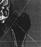
First scan age 77
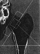
Second scan one year later
g/cm2
g/cm2
| First scan age 77 | Second scan one year later | |||||||
| REGION | BMD g/cm2 | T-score | Z-score | REGION | BMD g/cm2 | T-score | Z-score | |
| Neck | .688 | -2.4 | -0.3 | Neck | .654 | -2.7 | -0.6 | |
| Troch | .622 | -1.5 | 0.0 | Troch | .620 | -1.5 | 0.0 | |
| Total | .731 | -2.2 | -0.2 | Total | .731 | -2.2 | -0.2 | |
DEXA SCAN: (for the second scan):
The left femoral neck .654 gm/cm2 bone mineral density corresponds to osteoporosis and a T-score of -2.7. The left femoral neck bone mineral density has decreased approximately 4.9% since Lunar DEXA Scan previous date.
IMPRESSION:
Left femoral neck osteoporosis with approximately 4.9% decrease in bone mineral density since previous exam.
______________________________________
This was a 78-year-old woman who had serious degenerative joint disease including some scoliosis and disk disease in her spine. She had chronic back pain. Radiographs of the thoracic and lumbar spine showed no compression fractures. She had been taking estrogen. Another physician added alendronate for her back pain, but it caused GI distress so she wanted a second opinion. I suggested she did not need the alendronate but should stay on the estrogen.
A few months later her primary physician obtained this bone density report and was very concerned that she was getting so much worse and wanted to restart alendronate.
Updated 8/8/05

|
 |