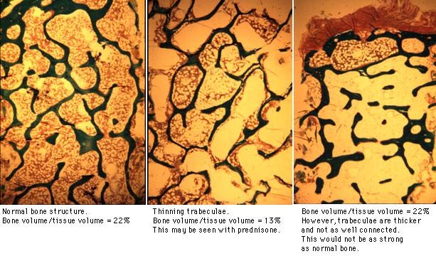
In addition to bone porosity, the bone strength is determined by the trabecular microstructure. Perforations of individual trabecula occur when resorption cavities are too deep. This, too, is seen with estrogen deficiency. The remaining trabecula are not as well connected and are mechanically weaker.

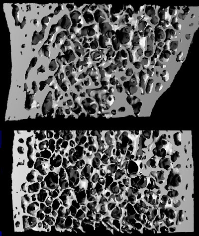
The diagram below shows that perforations weaken the bone, more than if the trabeculae just got thinner. The micro-CT scan is courtesy of Yebin Jiang, MD, PhD from University of California at San Francisco. It shows a paired iliac crest bone biopsy from one woman. The top is when she was 52.8 years old and premenopausal; the bottom was postmenopausal 5 years later. A series of these paired biopsies were analyzed and showed significant loss of trabecular micro-architecture.

|

|
Microfracture healing is another aspect of bone strength that is not measured by bone density. Trabeculae inside the bone may fracture and microcalluses are formed that resemble the calluses seen on xrays of long bones after a "macro-fracture". Osteoporotic bone is more susceptible to these fractures because the individual trabeculae do not have as many reinforcing connections. The calluses may represent a method of repairing the bone and even connecting some of the trabecula. Bone which has lost the ability to form these calluses will be weaker.
The age of the bone mineral crystals may also play a role in the strength of bone. This is an area that needs further research. Studies suggest that older bone is more brittle, and that one purpose of bone remodelling is to remove the old bone and replace it with newer, more elastic bone.
Mineralization density and biomechanics are discussed on the next page.
Updated 1/19/04
