Mammalian Digestive Systems |
|
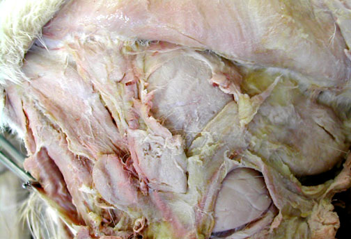
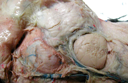
Cat Salivary Glands |
|
|
|
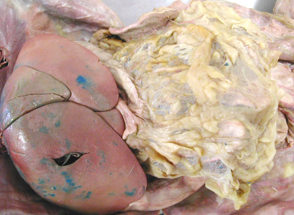
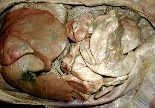
Cat "Mesenteries" |
|
The greater omentum is a double wall of peritoneal membrane & may act as a fat storage organ.
|
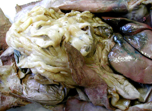
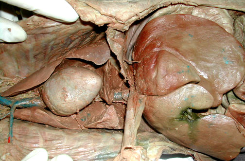
Cat liver & greater omentum. Find the gall bladder in a pocket of liver tissue.
|
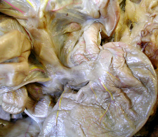
Another view of the greater omentum & the spleen which is really a cardiovascular organ.
|
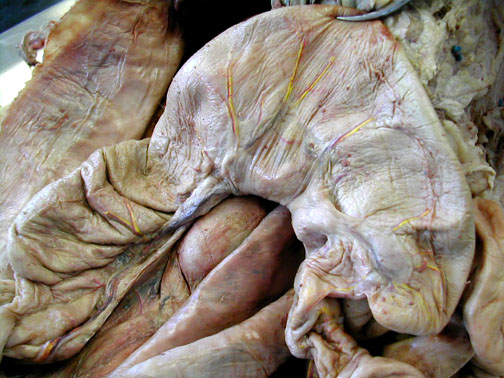
The falciform ligament is a remnant of the umbilical cord & attaches between the major right & left lobes of the liver. The falciform then attaches to the diaphragm (cut here). Heart & lungs are also visible.
|

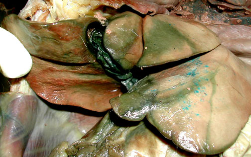
Cat: Abdominal Viscera |
|
|
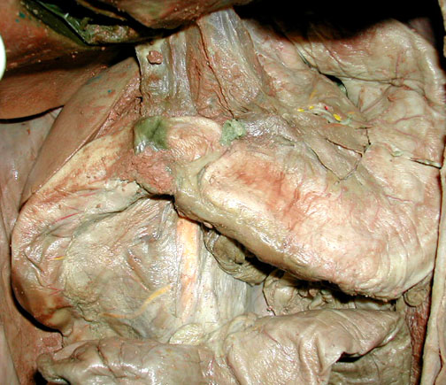
Leakage from the small, dark green gall bladder, stains surrounding portions of the liver green too.
|
|
|
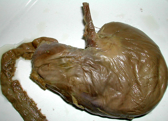
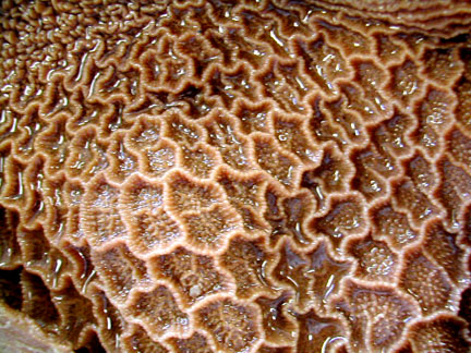
Stomach Exterior
|
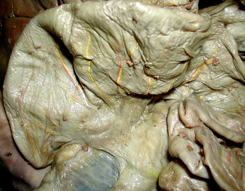
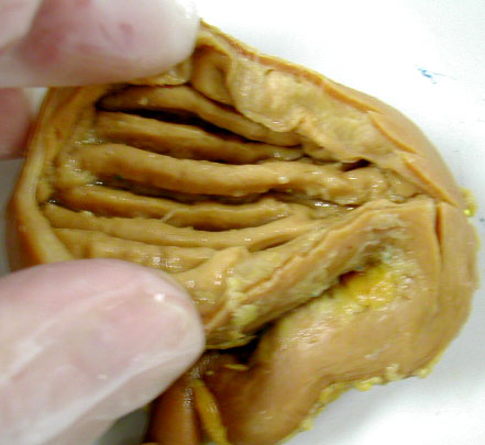
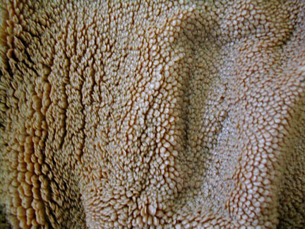
Stomach Rugae
|
Pyloric region of Stomach - with duodenum
|
Duodenum (first section of small intestine) with part of pancreas.
|
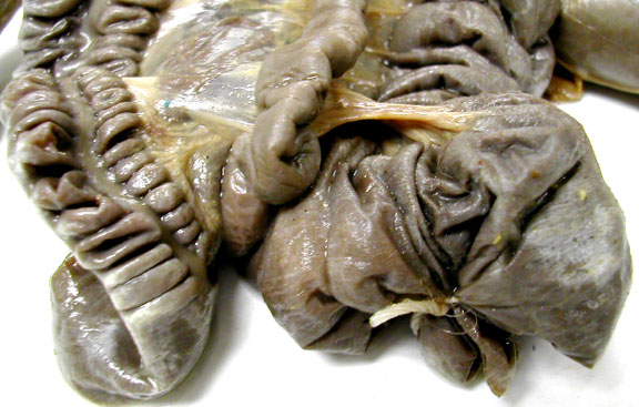
Ileocolic Junction has a sphincter controlling movement of materials from the small intestine into the colon.
|
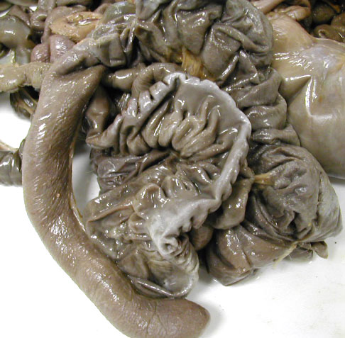
The cat's cecum is at the right & the colon extends in off to the left. The cecum of a cat is very small compared to that of a rabbit below.
|


Rabbit |
|
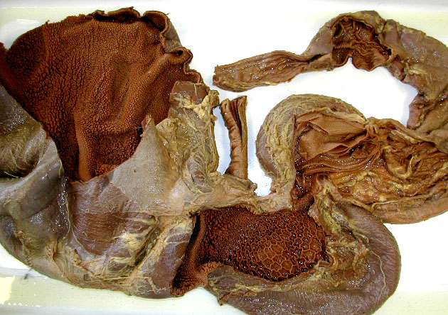
Stomach - with part of esophagus at the top & part of the small intestine & pancreas at the left. The stomach is large because green (moist) fecal pellets are reingested & stored here for a repeat of the digestive activity.
|
The rabbit cecum is a large, sac-like structure that ends with small 'appendix" on the left.
|
Small section of intestines - left - large intestine, middle - small intestine, right - cecum
|
|

| top of page |