|
Chondrichthyes: Dogfish Shark & Skate |
|
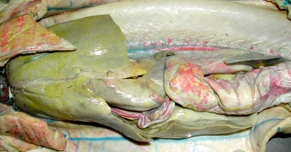
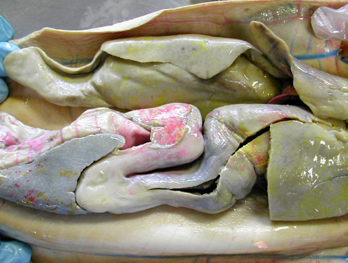
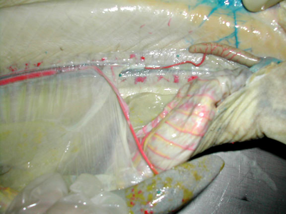
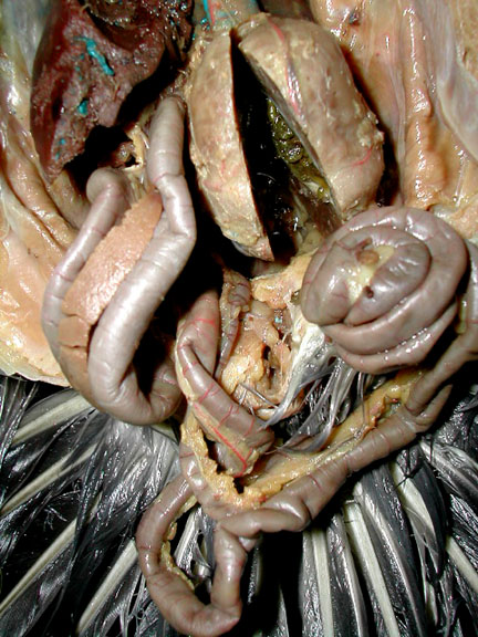
Overview of shark digestive system; large liver to the left.
|
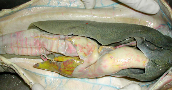
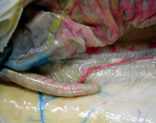
Another view of shark digestive system, the large liver is to the right.
|
|
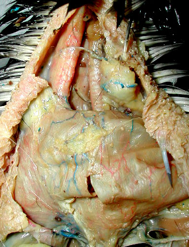
Another view of shark digestive system.
|
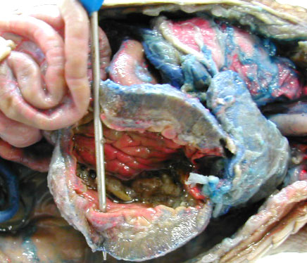
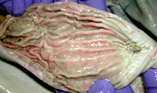
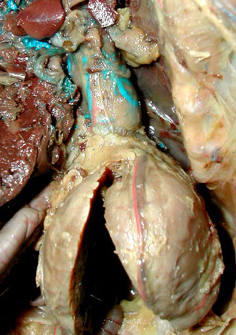
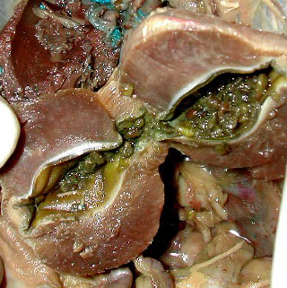
Fundic region of stomach opened to show rugae & esophageal papillae to the right.
|
|
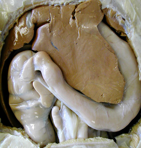
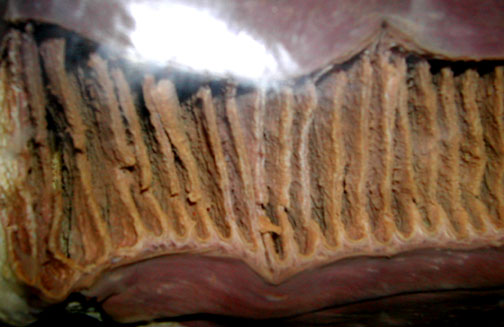
Opened section of spiral valve intestine
|
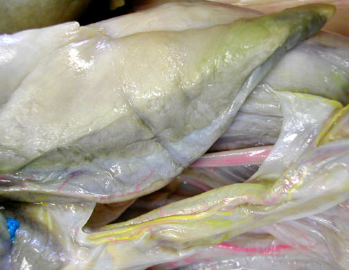
The gall bladder is the gray, flattened sac-like structure.
|
|
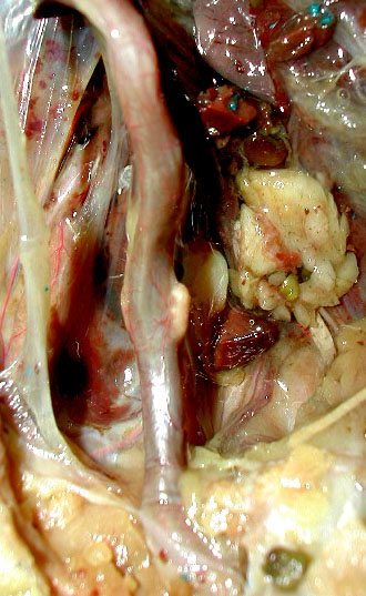
Shark rectal (digitiform) gland is at the far right, emptying excess salts into the colon.
|
Closer view of a shark rectal (digitiform) gland.
|
Aves: Pigeon |
|
This crop is a large, sac-like structure, with a small cut in it. Above the crop, the esophagus has been pulled to the left & the trachea (with it's cartilaginous rings is on the right.
|
A close-up look at the stomach with the smaller proventriculus above the cut gizzard.
|
|
Small intestines are below & to the left of the gizzard. The pancreas is the tan tissue between the 2 sections of the small intestine on the far left.
|
Ileocolic cecae are the tiny light tan structures along this part of the digestive tract & mark the boundary between the small intestine & the large intestine (colon).
|
|
The gizzard is opened to show it's thick muscular walls, keratinized lining & small gravel that birds swallow to help grind their food.
|