Mammalian Cardiovascular System Photos - part 2
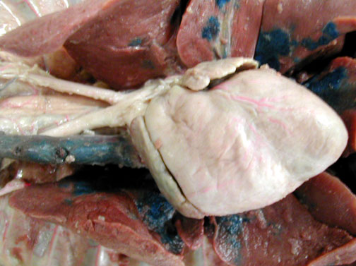
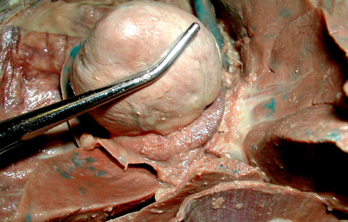
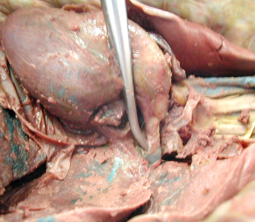
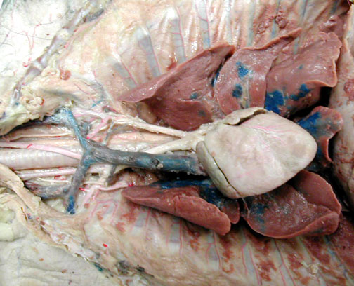
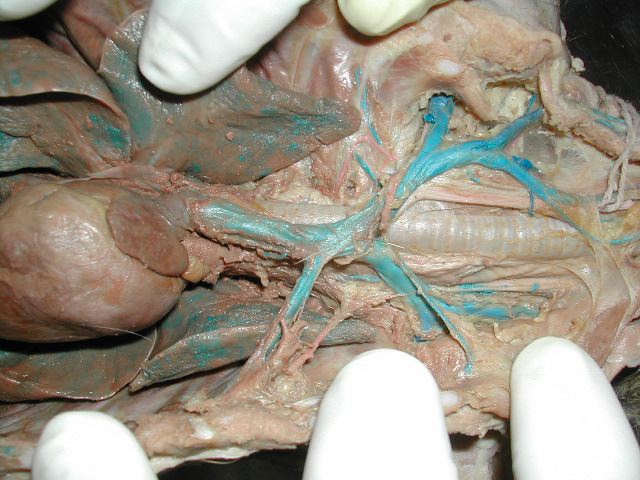
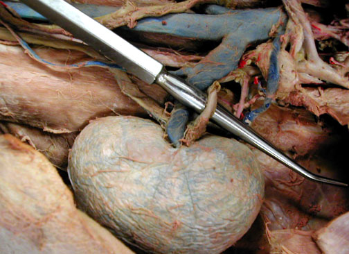
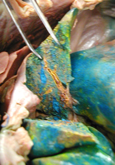
COMPARATIVE VERTEBRATE ANATOMY

Biology 453

Biol. 453 Home Page
Biol. 453 Lab Notes
Biol. 453 Photos
Biol. 453 Exam Page

Spleen

A normal healthy size.

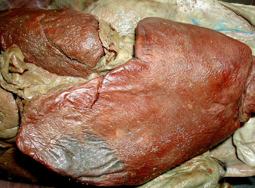
An unusually large size.


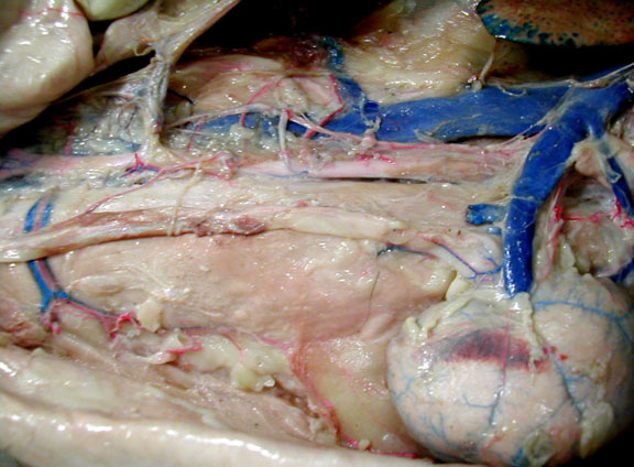
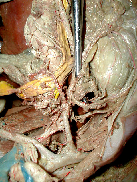
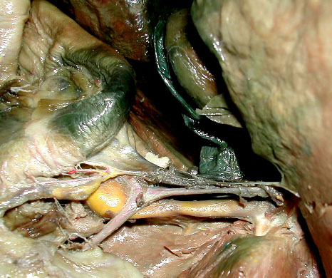
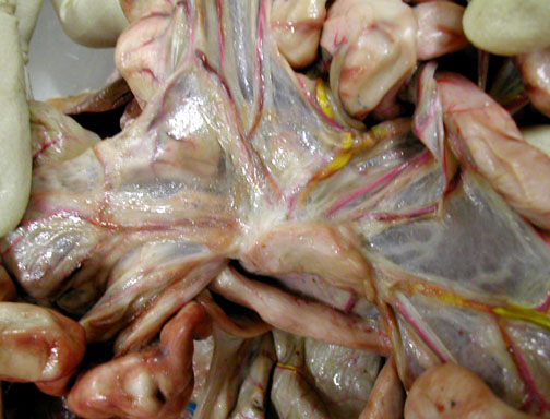
Cat Thoracic Vessels
Heart & Precava

Abnormal "Precava" - brachiocephalics don't join to form precava

Abnormal "precava" - left brachiocephalic attached along dorsal side of heart to enter R atrium

Abnormal "Post Cava" - "iliac" split occurs before renal veins branch off

Pulmonary Artery & Veins (left atrium was been damaged & part of it has been removed)

Pulmonary Artery & Veins


Cat: More Thoracic Region
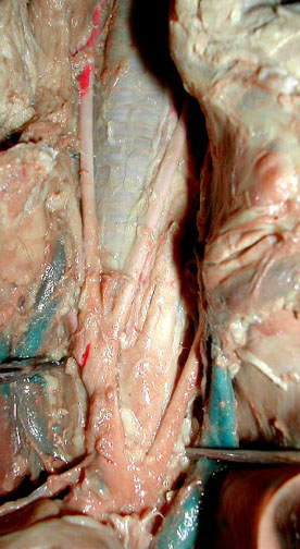
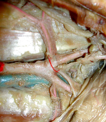
Arteries - brachiocephalic, R & L subclavians & R & L carotids
Veins - R & L brachiocephalic, R & L subclavians, R & L external jugular


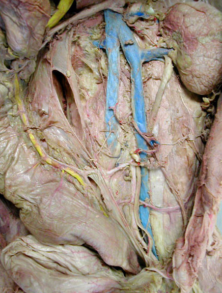
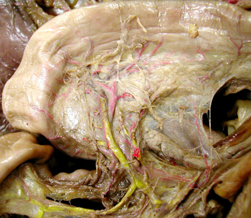
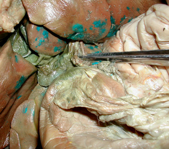
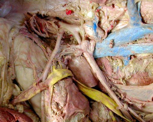
Cat Abdominal Region - Paired Parietal & Visceral Branches

Renal Artery & Vein

Renal vein

Normal Ileolumbar Artery & Vein - Tail & hind legs are to the left.

Ileolumbar A. & V. abnormally close to External Iliac A. - Tail & hind legs are to the right.


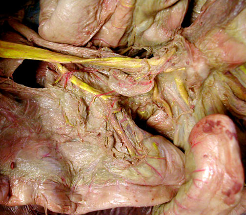
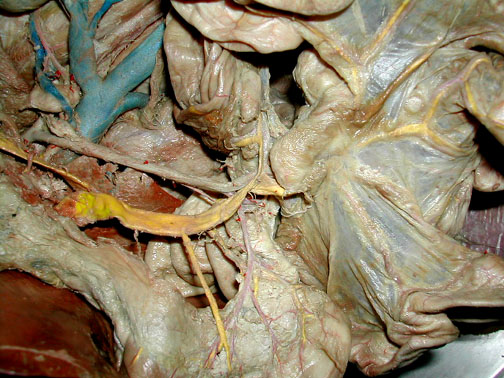
Cat Abdominal Region: Unpaired Arteries & Veins
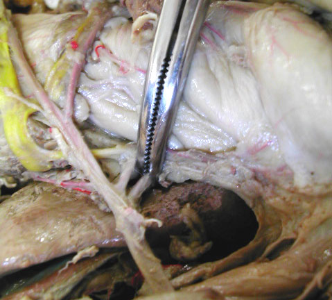
Celiac Artery & Its Branches

Posterior Panceaticoduodenal A. & V.
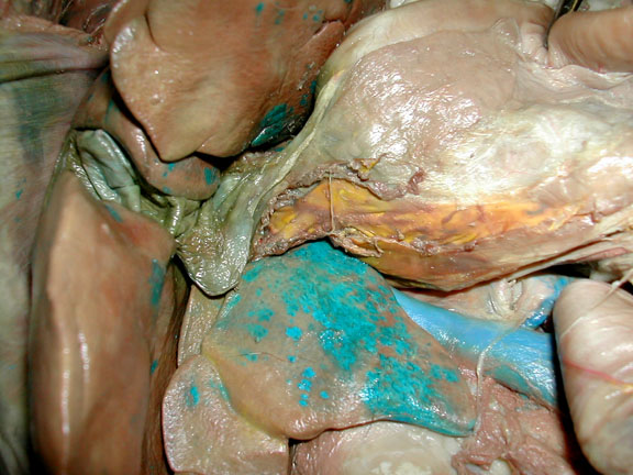
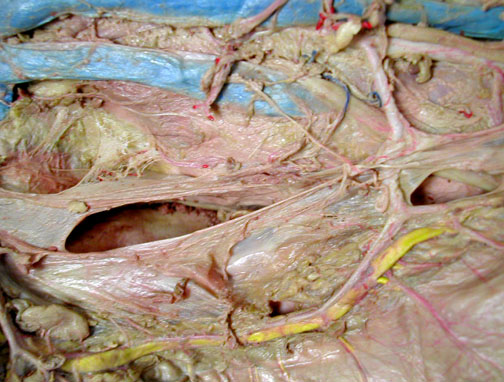
Hepatic A. & Hepatic Portal Vein - near liver

Branches of the hepatic portal vein (yellow) & hepatic vein (blue) within a cut section of the liver

Hepatic artery next to green bile duct. Gall bladder is above & to the left.

Hepatic portal vein (yellow) below bile duct (green).

Intestinal A. & V.

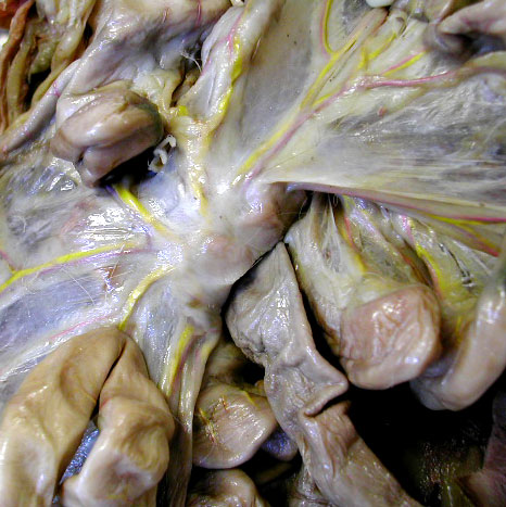
Intestinal veins are weakly stained in this cat.

Gastrosplenic Vein

Anterior Mesenteric Vein

Posterior Mesenteric Vein

| top of page |