SKELETAL MUSCLE PHOTOS - PART 1 CHONDRICHTHYES - Dogfish Shark |
|
Check out the variation between the different sharks & dissections. Click on each image to see the larger version.
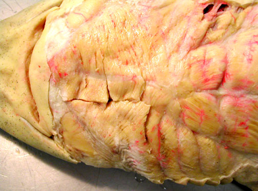
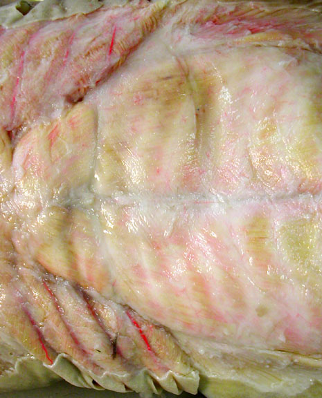
Red & white muscle fibers as seen in a transverse section through the tail. The red fibers are most readily visible on the right side, where you can see numerous red-stained blood vessels entering the tissue. Red fibers are aerobic, rich in myoglobin, giving it a darker color & rich in blood vessels to deliver oxygen. |
|
|
|


In the following photos, anterior is to the left or top, unless noted otherwise.
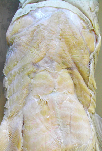
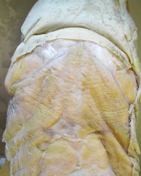
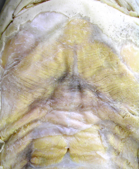
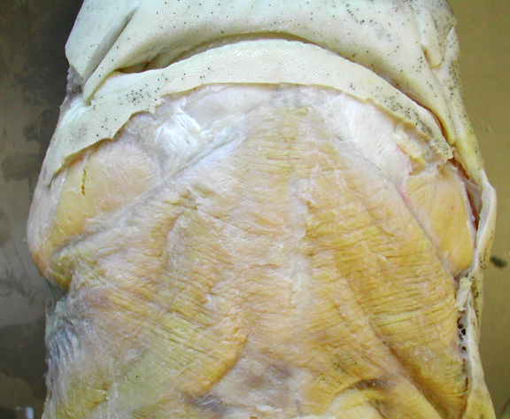
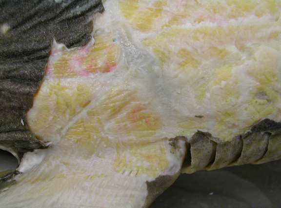
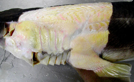
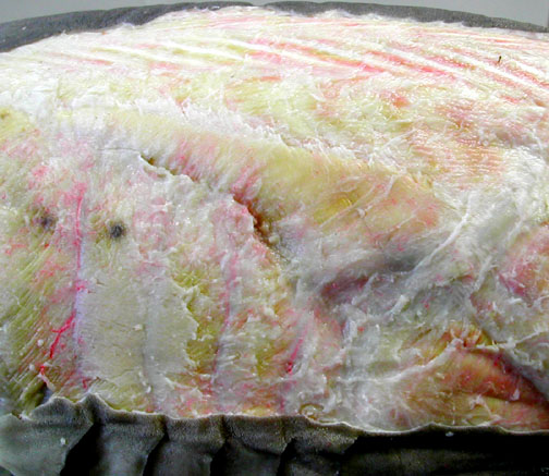
Ventral views of the superficial muscles show the throat to the pectoral fins. |
|

|
|
|
|
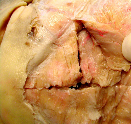
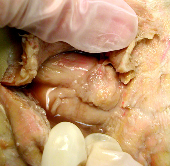
Closeup of the common coracoarcuals & intermandibularis. |
||
|
|
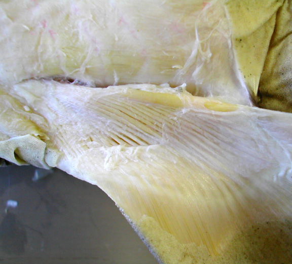
This view shows the coracoarcuals & the hypaxial muscles.
|
Closeup views of the intermandibularis muscle only. |
|

|
|
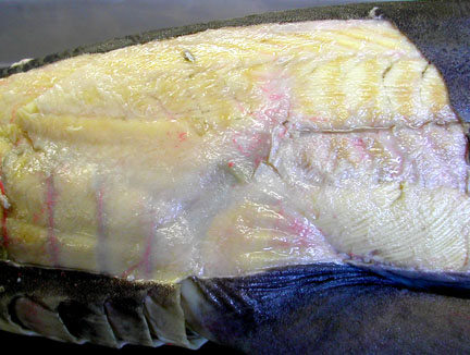
Views of the pectoral muscles |
|
|
|
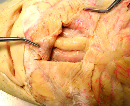
Ventral views with intermandibularis cut & reflected to reveal part of the interhyoideus. |
||
|
|
|
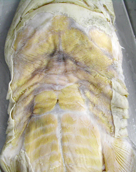
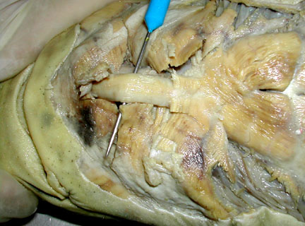
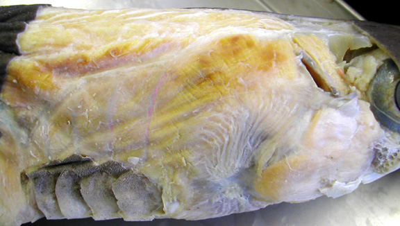
Ventral view of throat that expose the deep hypobranchial muscles - coracomandibular &/or coracohyoids. |
|
|
|

|
|
|
|
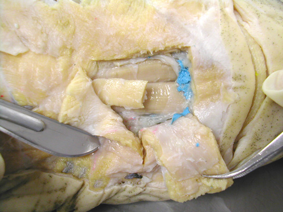
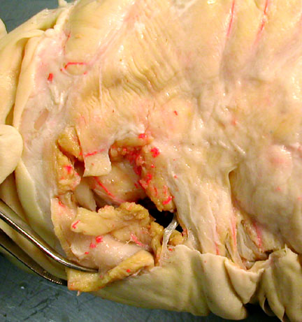
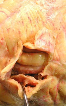
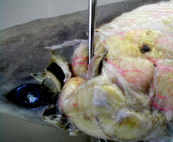
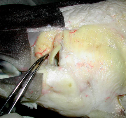
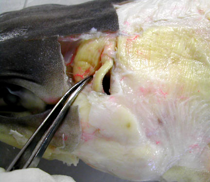
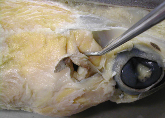
Lateral views of head with a probe inserted between spiracularis & levator palatoquadrati. Neurcranium is cut away to reveal these muscles. |
|
|
|
|
|
Lateral views of head show adductor mandibulae, levator hyomandibulae. |
|

|
|
Lateral views above gills that show cucullaris, dorsal constrictors, axial muscles & pectoral levators. |
|
|
|
|
|
| top of page |