Testudines (Chelonia)
|
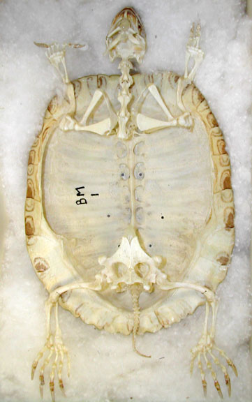
Turtle Skeleton -
Ventral view shows limbs, girdles & hyoid
apparatus.
|
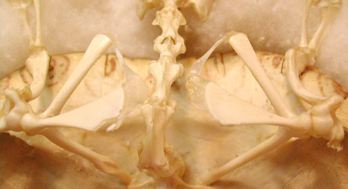
Close-up of pectoral
girdle has scapula, coracoid & clavicle.
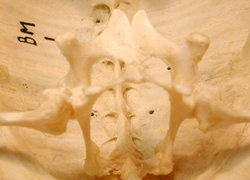
Close-up of the pelvic
girdle with pubis, ischium & ilium.
|
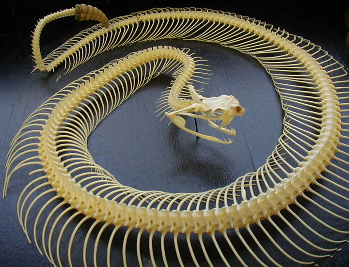
Lepidosauaria
|
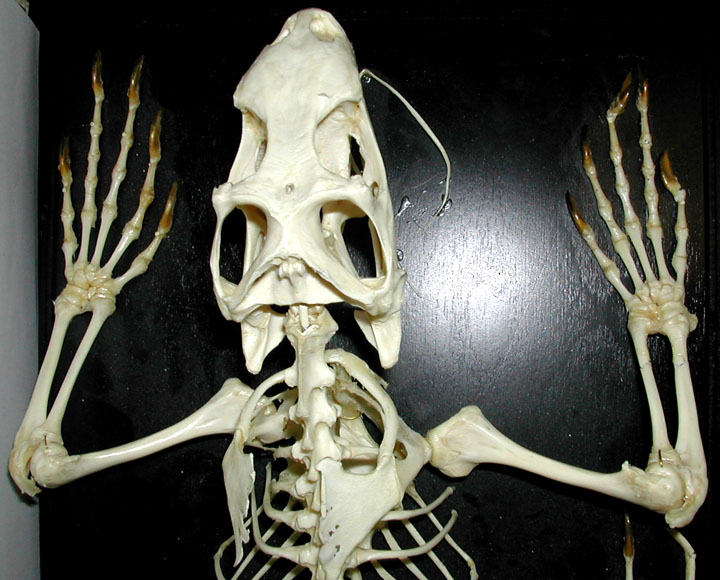
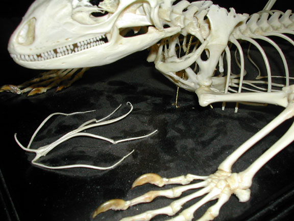
Rattlesnake
Skeleton
|
Iguana Skeleton -
dorsal view pectoral limbs
|
|
Iguana - pelvic girdle
& hind limbs

|
Iguana - sternum with a
bit of the interclavicle exposed medially.

|
|
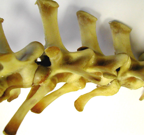
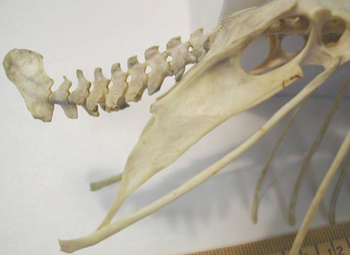
Varanid cervical
vertebrae, includes the axis at far left. The 1st
vertebra, the atlas is missing

|
Iguana Skeleton -
lateral view of the pectoral girdle & hyobranchial
apparatus
|
|
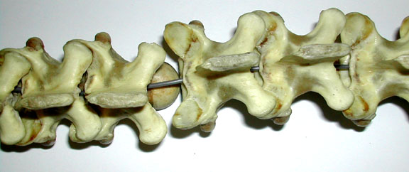
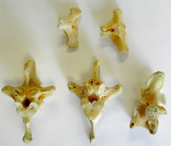
Varanid trunk vertebrae
(dorsal view). Locate the prezygapophyses.
|
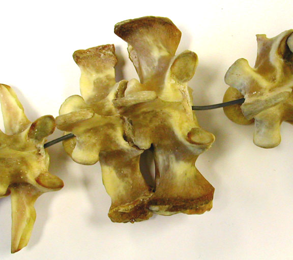
Varanid trunk vertebrae
(lateral view). Note the large articulation surfaces for the
ribs on the transverse processes.

|
|
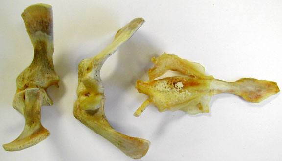
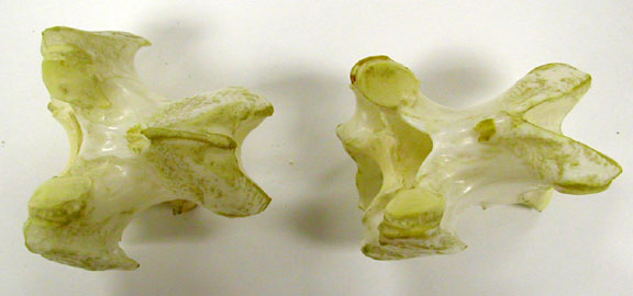
Varanid sacral
vertebrae (2).
|
Varanid caudal
vertebrae with large hemal canals within the "chevron"
bones.
|
Archosauria
|
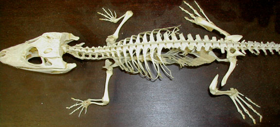
Alligator skeleton -
dorsal view.
|
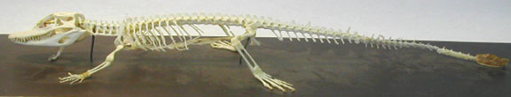
Alligator skeleton -
lateral view.
|
|
Alligator - pectoral
girdle bones with scapulae dorsal to the coracoids. The
interclavicle is at the right.
|
Alligator - gastralia
or abdominal ribs
|
|
Alligator paired humeri
at the left & paired femurs on the right.

|
Alligator lower limbs -
The radius & ulna pairs are at the left & the
tibia/fibula pairs are at the right along with the 2 largest
tarsal bones the astragalus & calcaneus.
|
|
Alligator vertebrae
& pelvis - Cervical, thoracic, lumbar vertebrae with
sacral vert. attached to ilium. Ischium bones face
posteriorly & the pubic bones are detached.

|
Alligator front &
hind feet - Parts of some digits are missing & the feet
show a few of the carpal or tarsal bones.

|
Aves
|
Gull Skeleton
|
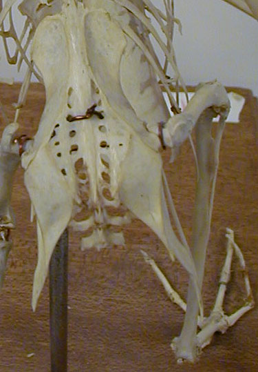
Gull Pelvis
|
|
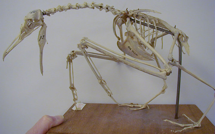
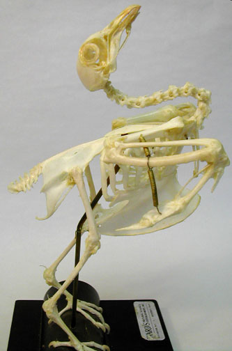
Pigeon Skeleton -
lateral view
|
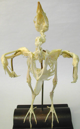
Pigeon Skeleton -
frontal view of the pectoral girdle & the wing.
|
|
Gull - frontal view of
the pectoral girdle & the wing.

|
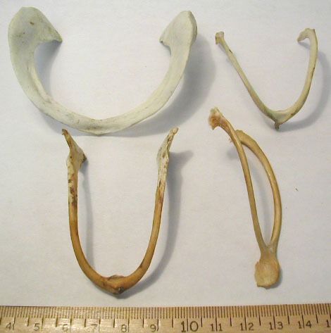
Miscellaneous bird
furculas (from the fusion of the clavicles &
interclavicles).
|
|
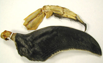
Humboldt Penguin wing
(below) & flattened forearm bones, radius, ulna,
carpometacarpus & phalanges (above).
|
Miscellanous
scapula-coracoids. The scapula angle upwards & are
thinner & longer than the coracoids.

|
|
Miscellanous bird
humeri (proximal heads to the left). The top 2 face
anteriorly & the bottom 3 face posteriorly.

|
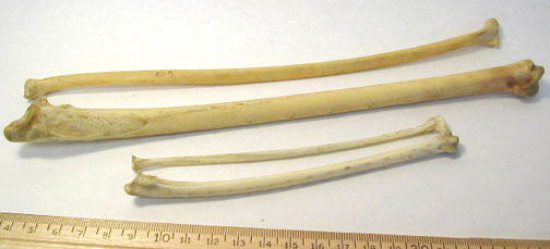
2 pairs of bird
radius-ulna bones.
|
|
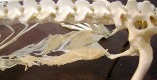
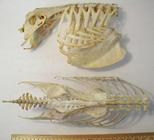
Bird "trunks" showing
ribs, synsacrum & pelvic girdles.
|
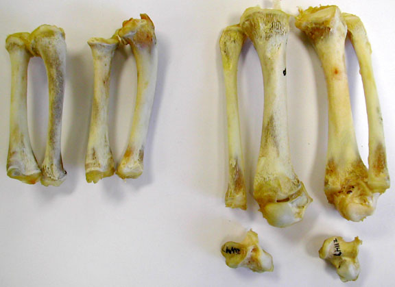
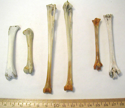
Bird leg bones - 2
unmatched femurs on the far left, paired tibiotarus &
fibulas in the center & 2 unmatched tarsometarsi on the
right.
|
|
Bird dorsal view of
cervical vertebrae showing the short neural spine &
pre-zygaphyses to the left.
Posterior view of cervical
vertebrae with transverse foramina, the heterocoelous
centrum & post-zygapophyses

|
Bird thoracic vertebrae
(lower)& caudal vertebrae (upper)
Caudal verterebrae ending in
pygostyle.
|
| top
of page |
