Click on each image to see a
larger version.
Cat (Eutherian) & Opossum (Metatherian)
Skeletons
|
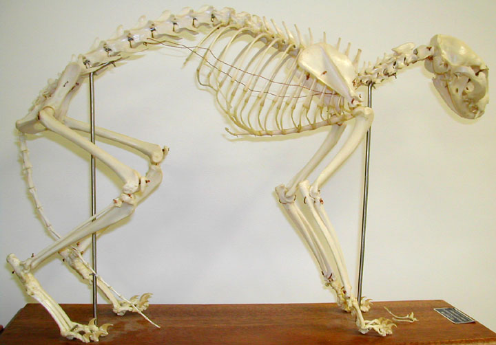
Cat skeleton lateral
view
|
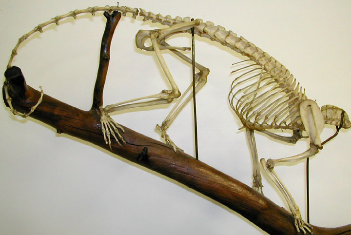
Opossum Skeleton
|
|
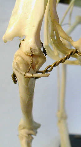
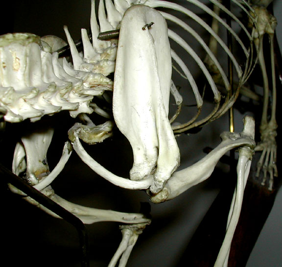
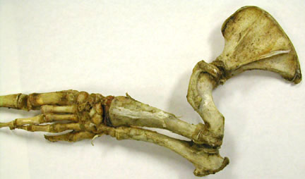
Cat hind limb

|
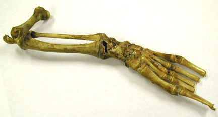
Opossum - note the
epipubic bones on pelvis and the tiny "chevron" bones
enclosing the hemal canals on the caudal vertebrae.
|
|
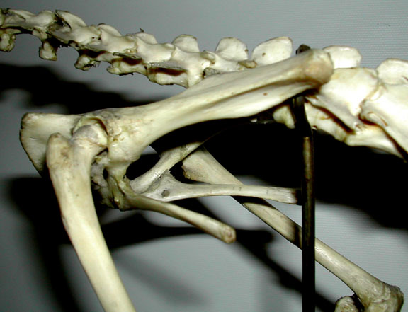
Cat - pectoral girdle
showing the "wired" in place, tiny clavicles. Cat clavicles
are suspended in muscle tissue normally.
|
Opossum - pectoral
girdle with large clavicles
|
|
Cat cervical
vertebrae
|
Cat thoracic
vertebrae

|
|
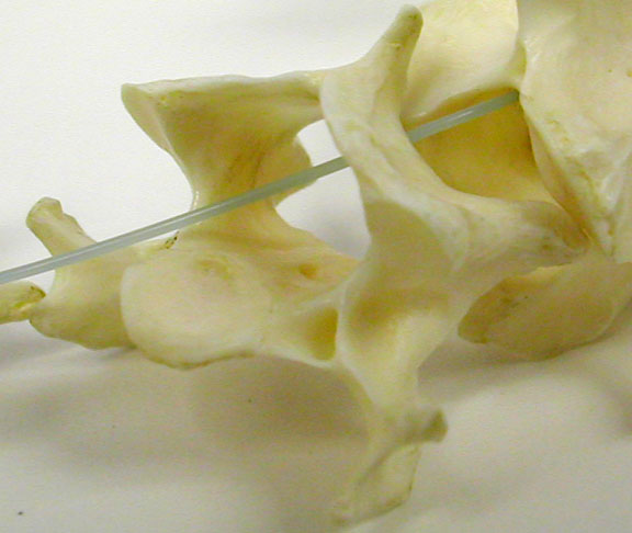
Cat lumbar & sacral
vertebrae

|
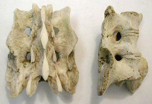
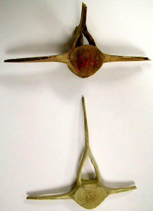
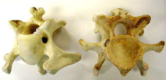
Porqupine sacral vert.
- dorsal view at left, ventral view at right.
|
|
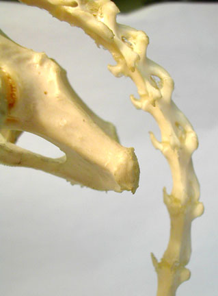
Cat caudal vertebrae
have very tiny hemal arches along the first part of the
tail.
|
Opossum caudal
vertebrae have hemal arches that sit ~ between the vertebrae
along the ventral side.

|
Human Skeleton
|
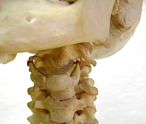
Human vertebrae -
showing atlas, axis above, thoracic vertebrae in lower left
& lumbar vertebrae in lower right.

|
Human skeleton with
cervical vertebrae & hyoid arch
|
|
Human clavicle &
scapula in position with the humerus.

|
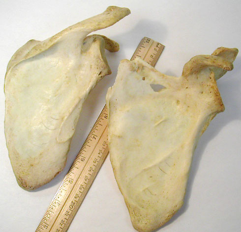
Human scapulae (lateral
view on the left & medial view on the right). Identify
the scapular spine, acromion process & coracoid
process.
|
|
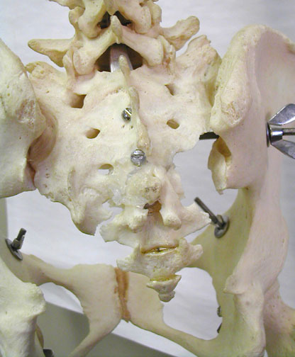
Human sacrum &
caudal vertebrae
|
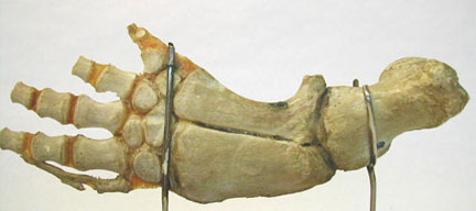
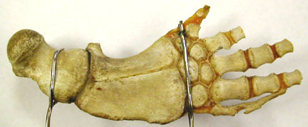
Human hand &
foot

|
Misc. Porqupine Bones
(Eutherian)
|
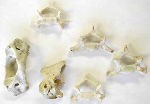
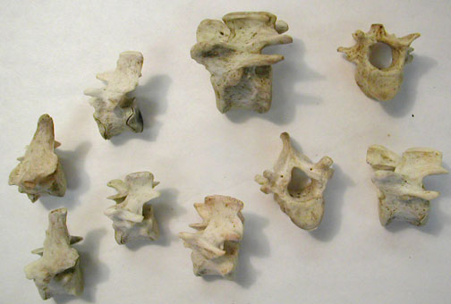
Cervical vertebrae -
lower left are the atlas & axis (it's odontoid process
is broken off, but note the large neural spine). The other
cervicals show their transverse foramina & are facing
anteriorly.
|
Thoracic vertebrae -
look for the rib attachments on the transverse processes
& centrum (not always easily visible on these small
vertebrae). The anterior sides of these vert. are facing
left.
|
|
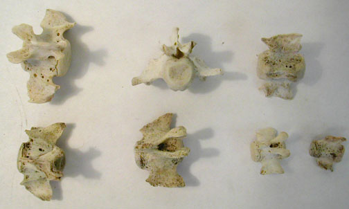
Lumbar vertebrae -
larger centra & no rib attachments. Anterior surfaces of
all vertebrae face forward or to the left. Find the pre
& post=zygapophyses.

|
Caudal Vertebrae have
tiny neural canals, reduced neural spines & transverse
processes, reduced zygapophyses. No hemal canals remain
attached, although they would have been present in these
vert.
|
|
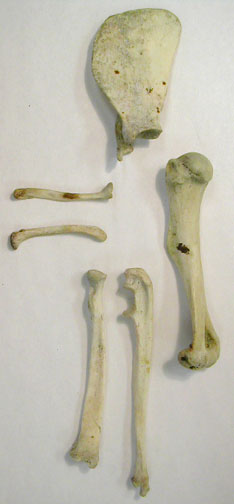
Front leg bones -
scapula (medial surface, so spine is hidden), clavicles,
humerus, radius & ulna.
|
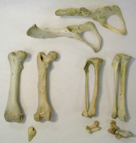
HInd leg bones -
"innominate", femur, patella (kneecap), tibia, fibula, a few
tarsals & phalanges.
|
Marine Adaptations
|
Dolphin front limb -
anterior (lateral) view
|
Dolphin front limb -
posterior (medial) view
|
|
Dolphin Vertebrae - The
upper vertebra is from the lumbar region & the lower,
narrow one is a cervical. Whales reduce the length of the
neck & limit neck movement.
|
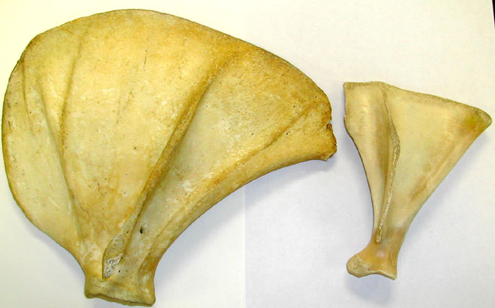
A large seal scaula on
the left & an ungulate scapula on the right.
|
|
Harbor Seal -
juvenile's forelimb
|
Harbor Seal -
juvenile's hindlimb
|
|
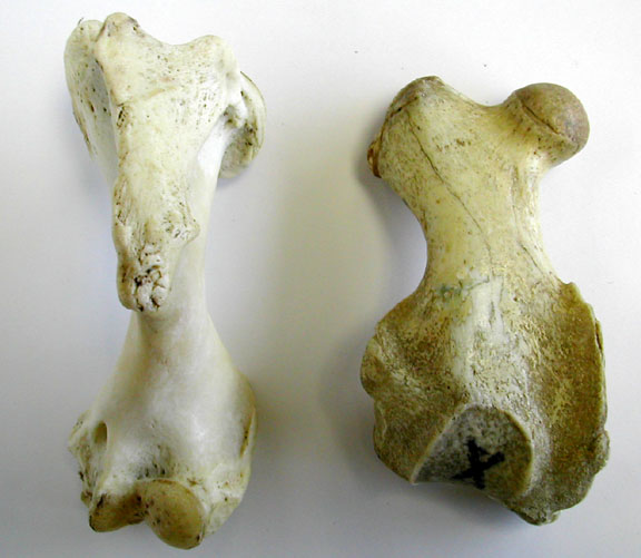
Harbor Seal - humerus
(left) & femur (right) anterior views
|
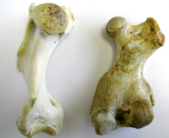
Harbor Seal - humerus
(left) & femur (right) posterior views
|
Flight Adaptations - Bats
|
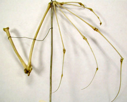
Megachiroptera - Bat
Wing
|
Close - up of that wing
to show the reduced ulna at the elbow.

|
Cursorial (Running) Adaptations of Large
Ungulates
|
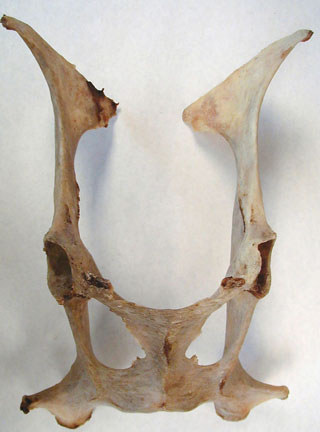
Springbok pelvic girdle
- ventral view with the ileum bones at the top, ischium at
the bottom & the pubic bones meeting in the
midline.
|
Miscellaneous ulnas -
lowest from a seal, largest is human & uppor ones show
the reduction typical of cursorial adaptions.
|
|
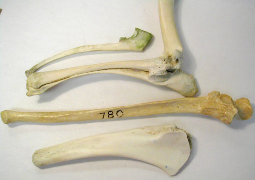
Anterior views of a
small ungulate (springbok) femur (left) & humerus
(right).

|
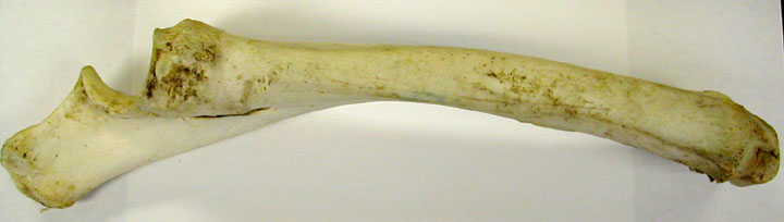
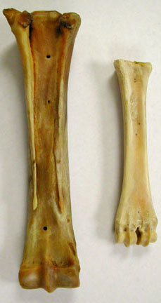
Horse radius & ulna
- note enlargement of radius & fusion of ulna to radius
that strengthens the leg & prevents rotation of the
foot.
Horse tibia

|
|
Horse & deer
metacarpals - posterior views. The deer metacarpal (right)
is made by the fusion of digits 2 & 3 & will connect
to two toes. The large horse metacarpal is from digit 3, but
you can see the reduced, # 2 & #4 metacarpals along
either side of # 3.
|
Horse phalanges, the
terminal phalange in enlarged to form the support for the
hoof.

|
|
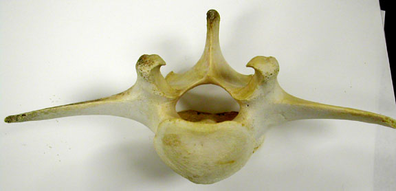
Atlas - 1st
cervical vertebra of large ungulate, anterior view.

|
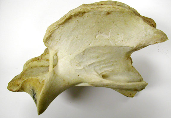
Axis - 2nd
cervical vertebra of large ungulate, lateral view.
|
|
Ungulate Cervicals -
left anterior & right posterior views
|
Lumbar vertebrae with
long transverse processes.
|
|
Lateral view of a
thoracic vertebrae from a large ungulate. Locate the
attachment points for the ribs on the lateral part of the
central & transverse processes. Find the
pre-zygapophyses.

|
Thoracic vertebae of a
large ungulate, posterior view showing the interlocking
post- zygapophyses.

|
| top
of page |
