|
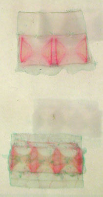
Vertebrae in plexiglass
- the red staining shows some ossification of the cartilage:
transverse sectioned above & sagittal sectioned
below

|
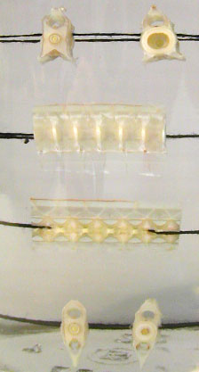
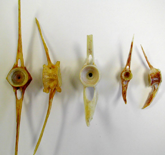
Vertebrae in fluid -
the 2 uppermost vertebrae are from the trunk region,
sectioned transversely to show different views of the
notochord in the interior. The lower 3 rows are views of
whole, sagittal sectioned & transversely sectioned
caudal vertebrae.
|
|
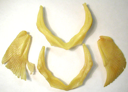
Pecotral girdle
(scapulocoracoid cartilage) & the left pectoral fin
showing basal & radial pterygiophores & brown
ceratotrichia.

|
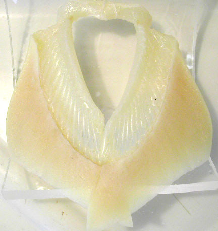
Shark - wax cast of
scapulocoracoid & pectoral fin ptyergiophores; top is
anterior view of scapulocoracoid, lower is posterior view of
scapulocoracoid
|
|
Males have pelvic
fins with claspers

|
Females have simpler
pelvic fins.
|
|
Amia Skeleton -
anterior portion of skeleton

|
Amia - posterior
portion of skeleton with damaged tail.

|
|
Amia pectoral girdle
shown ventrally. Note the large, bony cleithrum & small
scapulacoracoid cartilage that holds the pectoral fin.
Locate the small row of pterygiophores at the base of the
fins.
|
Amia - pelvic girdle,
ischiopubic plates float in muscle tissue
|
|
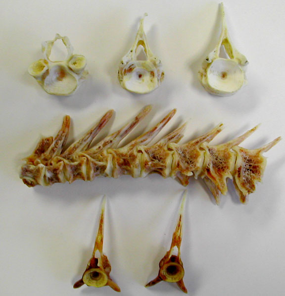
Trunk Vertebrae -
diverse views
|
Caudal Vertebrae -
Hemal canals on the ventral side of the centrum are usually
larger than the neural canals.
|
|
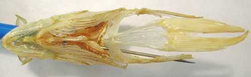
Perch Skeleton

|
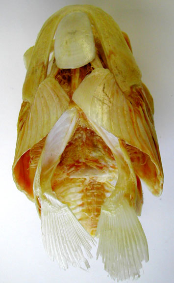
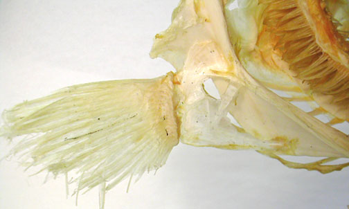
Perch ventral view of
jaws, hyoid & pelvic girdle (made of ischiopubic pates)
& pelvic fins.
|
|
Cod pectoral girdle
bones, part of the large cleithrum on the left side &
the scapula (dorsal) & coracoid (ventral) that hold the
fin.
|
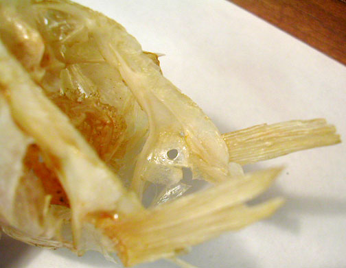
Perch Lateral view of
skull. Along the ventral line, note the jaws, hyoid arch,
part of the cliethrum & scapula of the pectoral girdle
(the coracoid may be missing here). The pelvic girdle & its fins attach to the cleithra.

|
|
Perch - angled,
interior view of pectoral girdle showing cleithrum, scapula & coracoids.
|
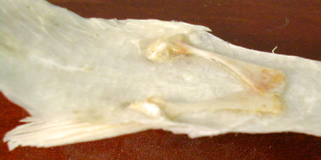
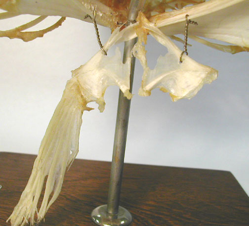
Cod pelvic girdle made
of ischiopubic plates that normally attach to the pectoral
girdle at the midline (cleithra).
|
|
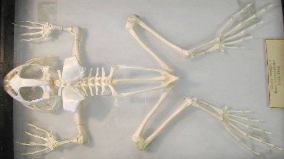
Frog Skeleton - Dorsal
View
|
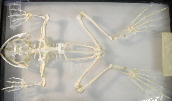
Frog Skeleton - Ventral
View
|
|
Frog vertebral column & pelvis, dorsal view (top) & ventral view
(bottom).

|
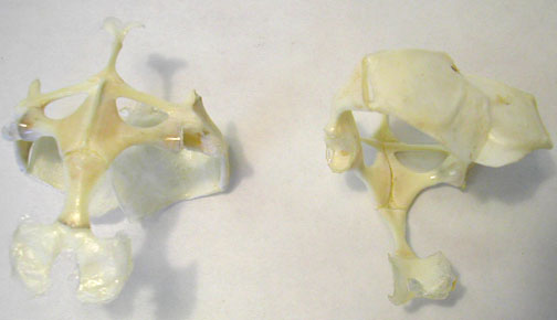
Frog pectoral girldes-
on the left is a ventral view of pectoral girdle showing
sternal elements, clavicles & coracoids. & on the
left is a dorsal view of the suprascapulae.
|
|
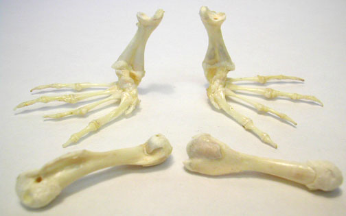
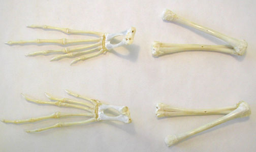
Frog - semi-articulated
front limbs
|
Frog - semi-articulated
hind limbs
|
|
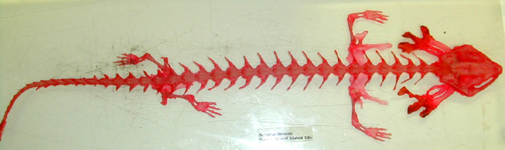
Necturus Skeleton -
Dorsal View
|
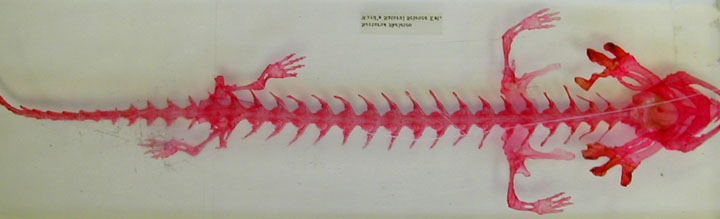
Necturus Skeleton -
Ventral View
|
|
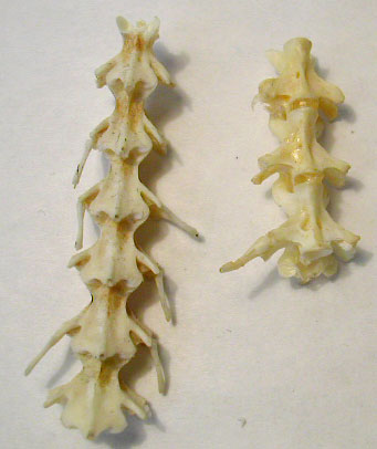
Necturus vertebrae -
Left section shows the dorsal side of vertebrae &
exposes 1 set of pre-zygapophyses at the top) & in the
right section, the vertebrae are upside-down to see 1 pair
of post-zygapophysis facets (at bottom).
|
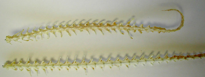
Necturus vertebral
columns - the upper one is the ventral view & lower one
shows dorsal view. Upper column has clearly recognizable
sacral vertebra. Both show the atlas (1st & only cervical) vertebra.
|
|
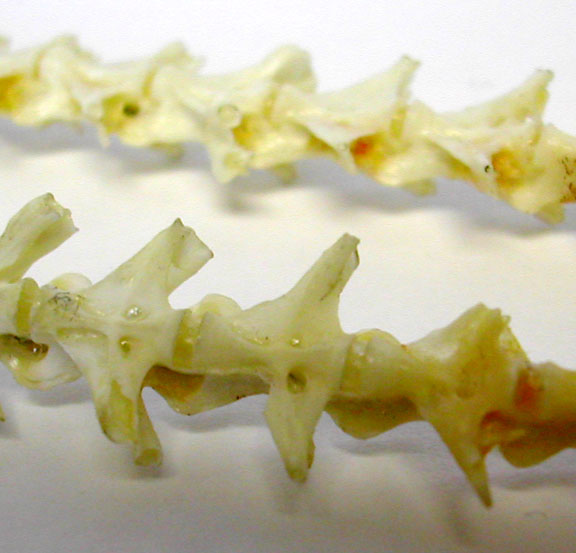
Necturus showing
single, sacral verteba with enlarged transverse processes & rib still present.
|
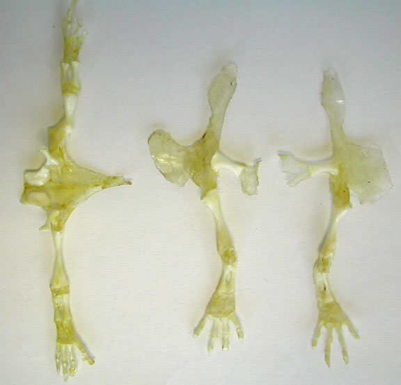
Necturus - dried front
(right) & hind limbs (left). Both have girdles
attached.
|


