|
Hagfish - note
the row of ducts located ventrally along the body for the
exit of mucous from the numerous slime glands

|
Hagfish - this
row of larger pores are the exits for water from the
hagfish's gills.

|
|
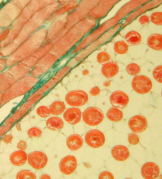
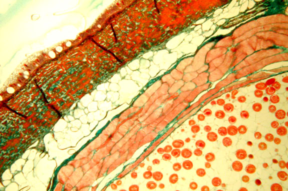
Hagfish Skin -
higher power shows thin epidermis with unicellular
mucous glands (at bottom of image), thick dermis with
connective tissues (green) & smooth muscle (red).
Chromatophores are numerous at the epidermal & dermal
boundary.

|
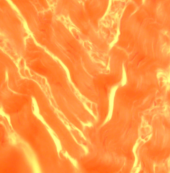
Hagfish Slime Gland
- high power shows part of large, deep epidermal slime
gland (at bottom) & surrounding skeletal muscle that
must contract to expel the slime.
|
|
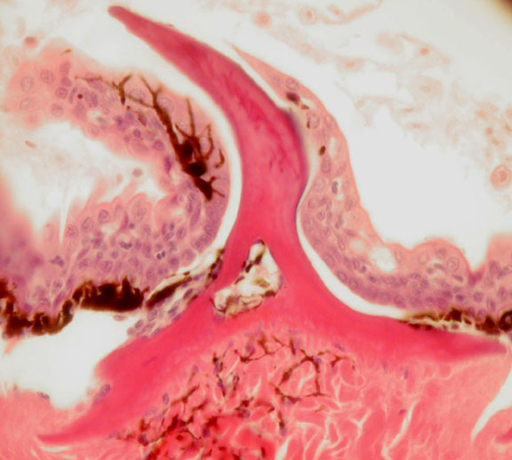
Lamprey Skin -
shows epidermis at far right with some unicellular mucous
glands. Dermis is relatively thin compared to the hagfish.
Far left shows some muscle tissue in cross section.
|
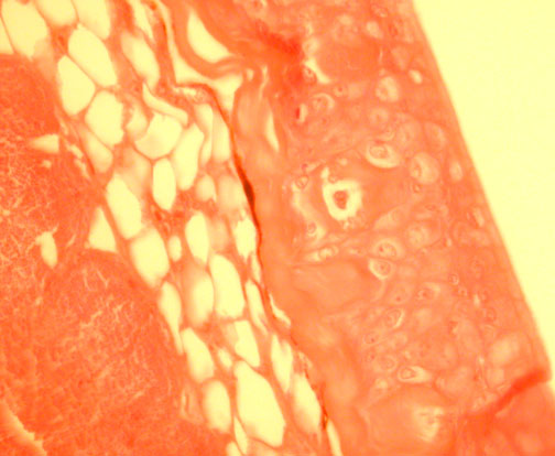
Hagfish Skin - Low
power shows epidermis & dermis of skin & the
skeletal muscle that surrounds slime gland.
|