Click on each image to see a
larger version.
MAMMALIA -
CAT
|
Cat - dorsal
view

|
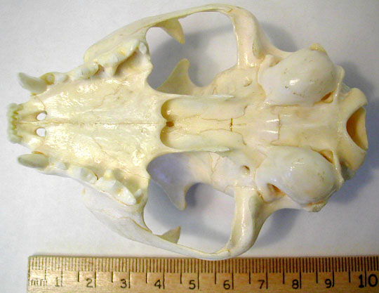
Cat - ventral
view
|
|
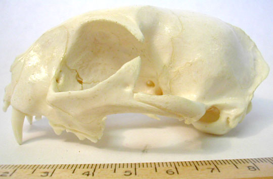
Cat - lateral
view
|
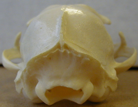
Cat - posterior
view
|
|
Cat sagittal
views - showing the turbinates (nasal conchae) on the
left & the ethmoid plate on the right.
|
Cat lower jaw -
find the coronoid, angular & condyloid processes.

|
|
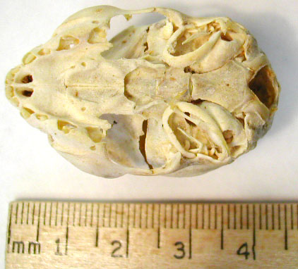
Youngest Kitten
Skull - shows the angular ring (dermatocranium) with
the malleus (splanchnocranium) inside the ring. The tympanic
bulla (new dermatocranium bone) is the small round bump
partly underneath & posterior to the angular
ring.
|
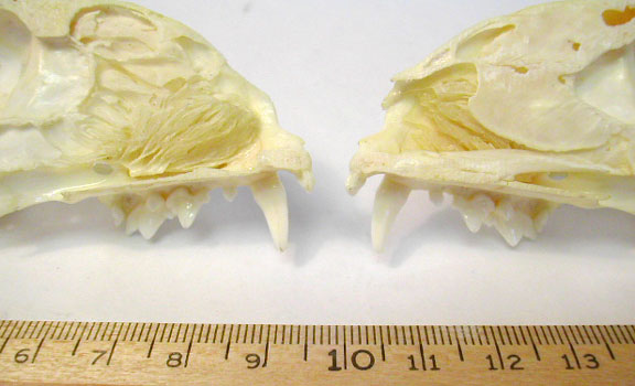
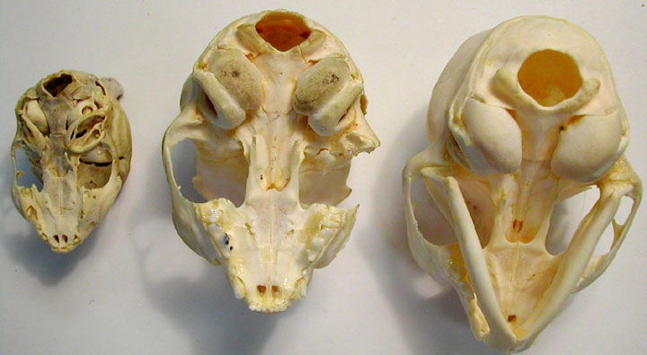
Kitten Skulls -
series shows growth of tympanic bulla & it's fusion with
the angular ring.
|
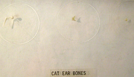
Cat Ear Ossicles
- malleus - far
left
- incus -
center
- stapes -
right
|
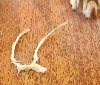
Cat Hyoid arch -
not in correct anatomical position obviously!
|
MAMMALIA - HUMAN
|
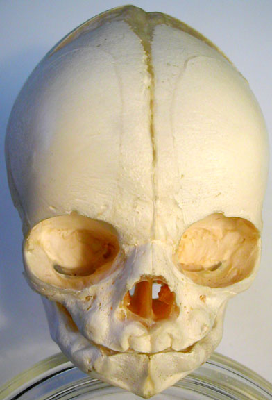
Neonate Human
Skull - anterior view, you can still just detect the
different origins of the premaxilla & maxilla which will
be completely fused in adults.
|
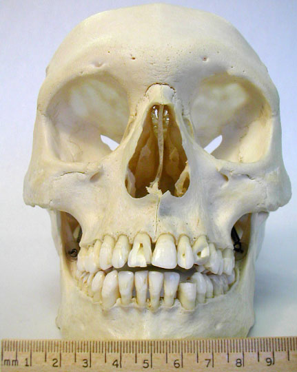
Adult Human
Skull - anterior view
|
|
Neonate Human
Skull - lateral view

|
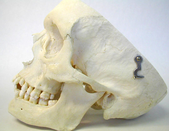
Adult Human
Skull - lateral view
|
|
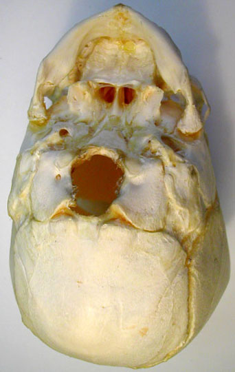
Neonate Human
Skull - ventral view shows the separate occipital
bones
|
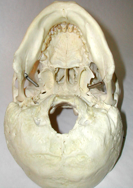
Adult Human
Skull - ventral view
|
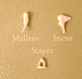
Human Ear
Ossicles - homologies below
- malleus -
1st arch splanchnocranium =
articular
- incus -
1st arch splanchnocranium =
quadrate
- stapes -
2nd arch splanchnocranium = columella =
hyomandibular
|
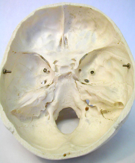
Adult Human Skull
- interior view
|
| top
of page |
