Click on each image to see a
larger version.
Cat Urinary System |
|
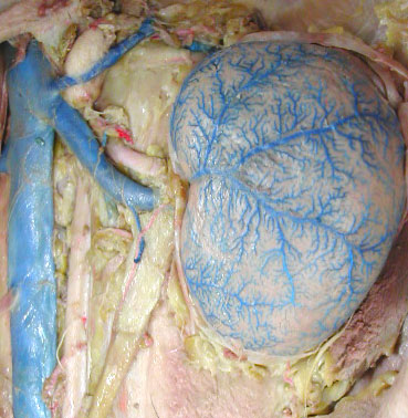
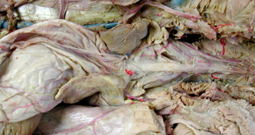
Cat kidney with ureter
exiting to the right
|
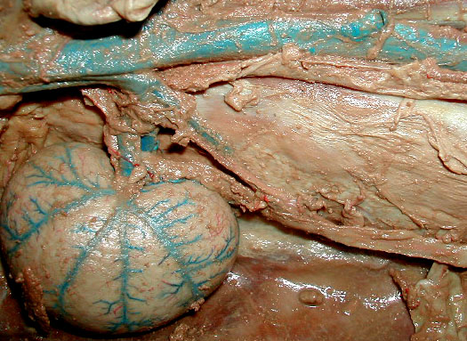
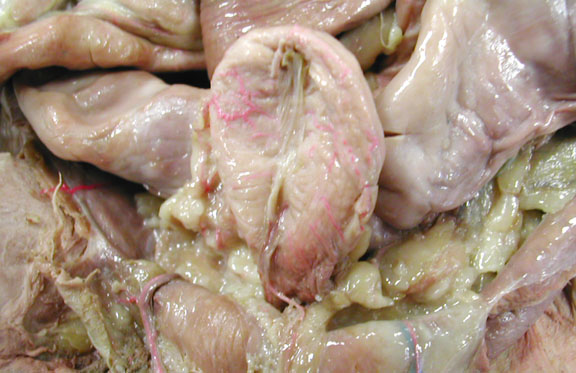
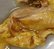
Cat kidney with visible
adrenal gland, ureter, renal artery & renal vein
|
|
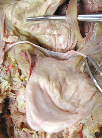
Cat kidney sectioned;
identify renal cortex, renal medulla & (small) renal
pelvis.

|
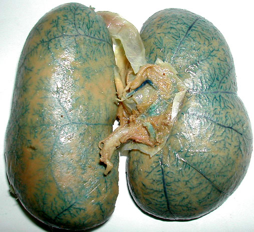
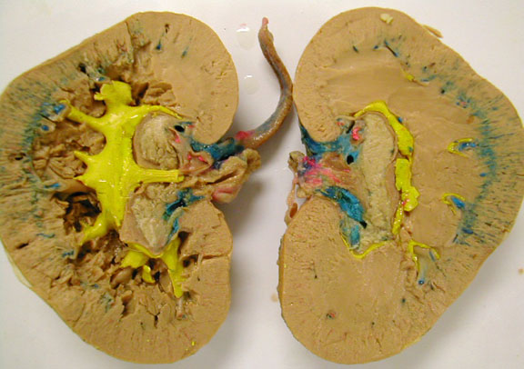
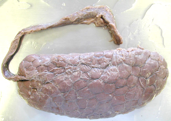
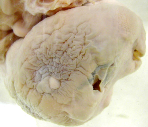
Abnormally large,
single kidney (no kidney formed on other side of
body).
|
|
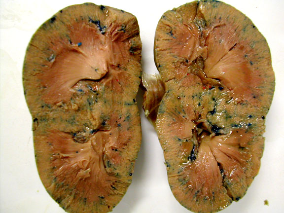
Abnormal Kidney after sectioning shows has 2 renal pyramids.
|
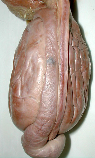
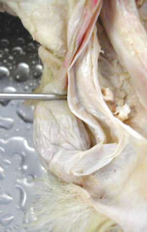
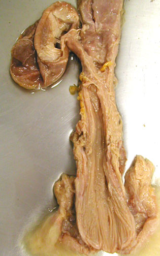
Male cat's urethra
extending to the right, with stretched bladder to the
left
|
|
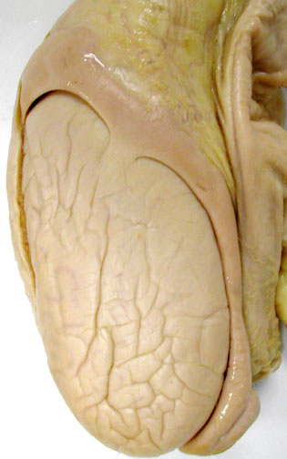
Female cat urinary
bladder (slightly enlarged uterus extends L & R behind
bladder)
|
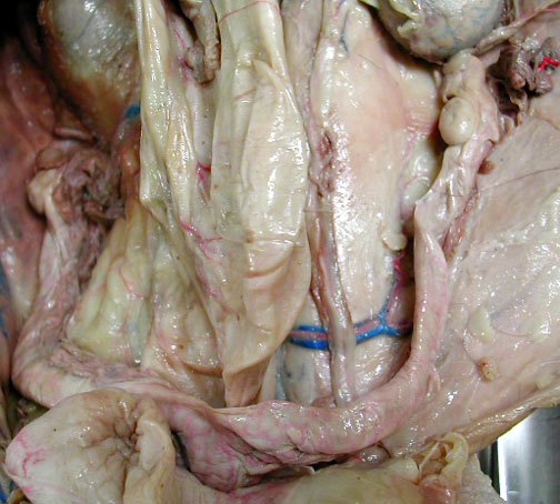
Male cat - dorsal side
of urinary bladder, ureter (crossing over blunt probe) &
vas deferens (white ducts, on R & L, one is held by
forceps)
|
Miscellaneous Mammalian Kidneys |
|
Sheep Kidney
|
Rabbit Kidney

|
|
Dall Porpoise KIdney (lobular design) & ureter
|
|
Male Reproductive System: Bull |
|
Tunica albuginea pulled
away from part of the testes

|
Cremaster muscle
(skeletal muscle from the external obliques) relaxation
moves scrotum further from body when testes are too
warm)

|
|
Testis & vas
deferens is the narrow duct on the left.

|
Pampiniform plexus
(cardiovascular countercurrent heat exchanger in spermatic
cord)

|
|
Epididymis (left) & vas deferens (thinner tube on the right) on the
testes
|
Testis & epididymis
|
Male Reproductive System: Cat |
|
Cat testes &
epididymis visible on (upper) L side & testes & vas
deferens visible on (lower) R side. Note the pampiniform
plexus readily visible next to the vas deferens.
|
Cat scrotum opened with
testes, vas deferens & tunica albubinea visible
|
|
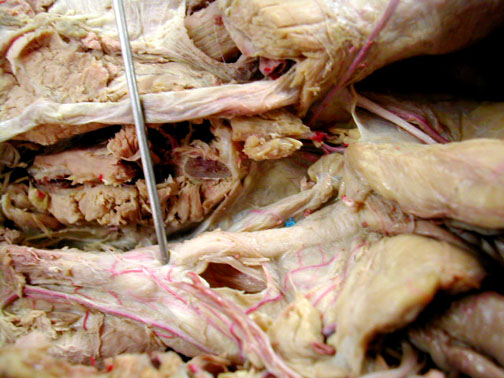
Cat prostate gland
(just below & to the left of the pin insertion. The
spermatic cord is the whitish cylinder above the
urethra.
|
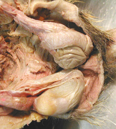
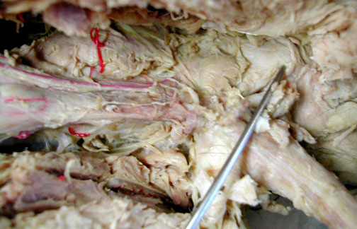
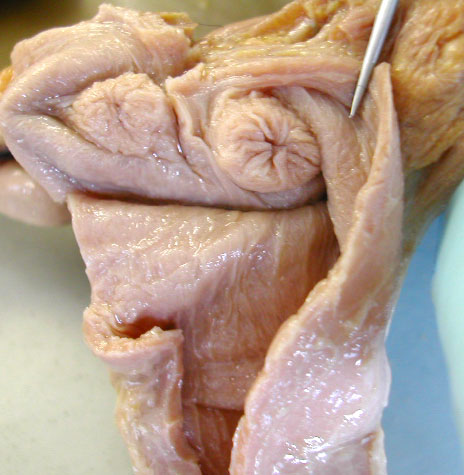
Cat bulbourethral gland
(the 2 round structures just to the left of the
probe).
|
|
|
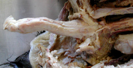
Cat penis
|
Female Reproductive System & Embryos: Pig |
|
Ovary hidden within
infundibulum of Fallopian/uterine tube

|
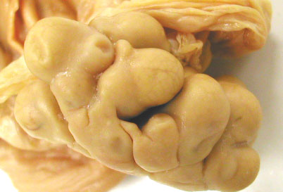
Ovary - at ovulation, 1
rupture & 1 egg ready
|
|
Ovary - entire (the
large round masses within it are probably corpora
lutea
|
Bisected ovary with at
least 2 large corpora lutea (far R & L of probe
tip).

|
|
Uterus - early
pregnancy, cervix is at the bottom, center
|
Pig uterus -
Fallopian/uterine tube leading into uterus
|
|
Early embryos, 1 has
amniotic sac around it. The liver is the large grey mass in
the mid-ventral part of the body.

|
Larger embryo
with diffuse placenta
|
Female Reproductive System & Embryos: Goat |
|
Bicornuate uterus -
pregnant
|
Cotyledonary
placenta

|
|
Uterine cervix
|
Ovary on Uterine
wall
|
Female Reproductive System: Rabbit |
|
Female repro.
tract
|
Urogenital sinus - with
vagina & necrotic bladder
|
|
Ovary is the small,
bean shaped mass in the center.
|
Paired cervices in
duplex uterus
|
Female Reproductive System: Non-pregnant Cat |
|
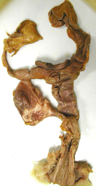
Uterine Horn
|
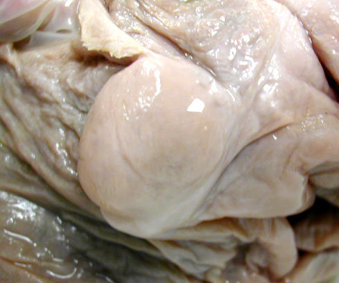
Ovary & Fallopian tube

|
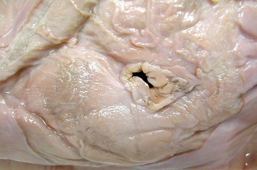
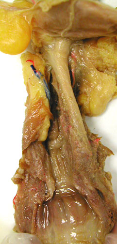
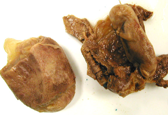
Urogenital sinus |
The probe is hear the exit of urine from the urethra. Below that, is the common exit for urinary & reproductive systems the urigenital sinus.
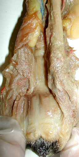
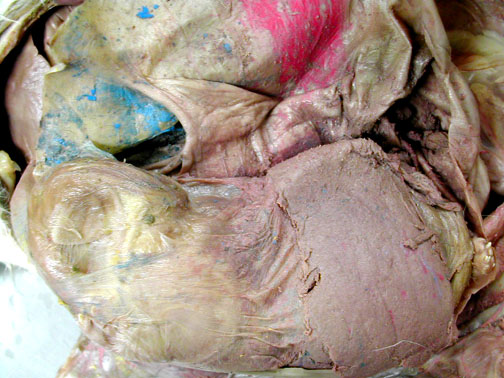
|
The urethra is the narrow tube running along the right. Part of the vagina is cut open on the left. The larger, darker space at the bottom is the urogenital sinus.
|
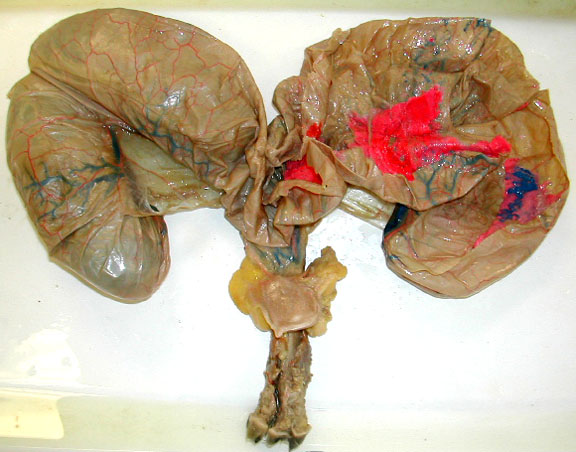
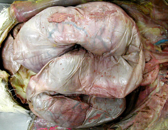
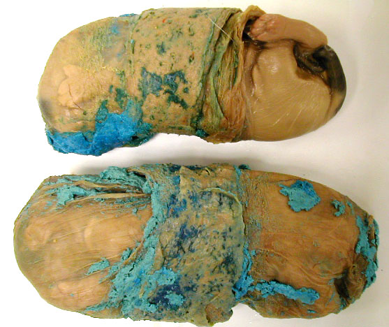
Female Reproductive System: Pregnant Cat |
|
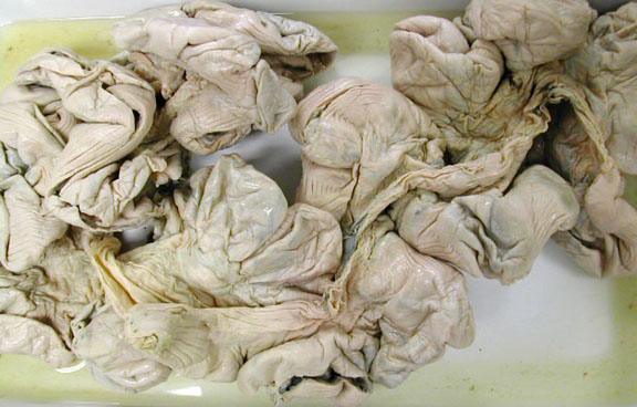
Pregnant female reproductive tract & urinary bladder removed from the body.
|
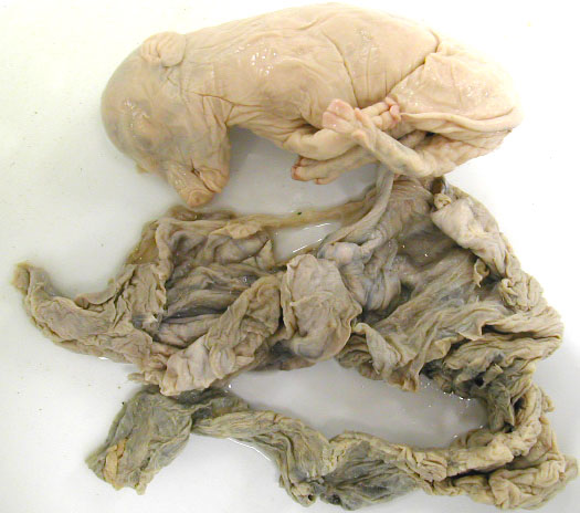
Near term uterus fills much of the abdominal cavity. She had 5? kittens.
|
|
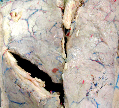
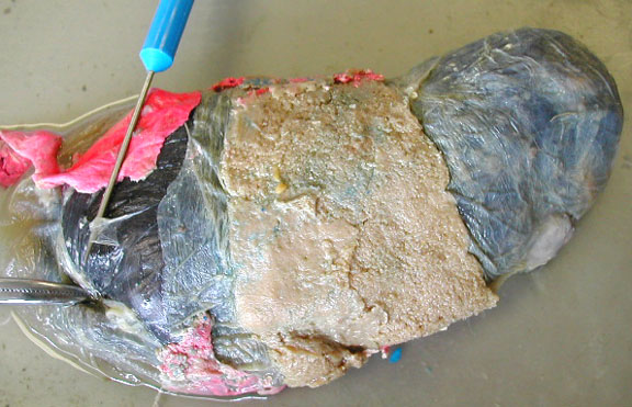
Early pregnancy - uterus shows enlarged compartments for 4 embryos.
|
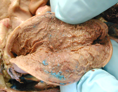
Interior of uterus showing zonary placentae region (the blue latex marks the veins going to it) This may be a post-partum uterus? or very early pregnancy stage.
|
The mammary glands rund from the thoracic region to the pelvis.
|
A closer view of mammary tissue.
|
Female Reproductive System: Embryo, Placentas & Extraembryonic Membranes |
|
Early embryo - amnion still around embryo, placenta opened
|
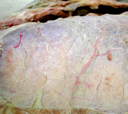
Zonary placentae - upper one has part of chorioallantois torn to reveal amnion
|
|
Early zonary placentae
|
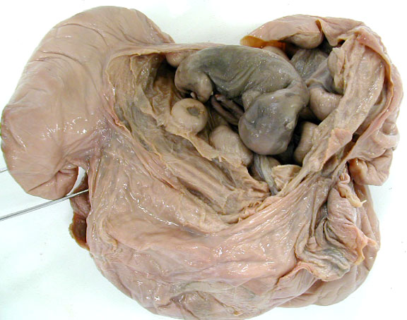
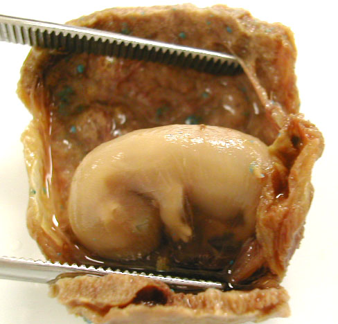
Kitten in uterine horn with zonary placenta partly peeled from uterine wall
|
|
Kitten has chorionallantois partly removed on the left by the forceps with blue probe lifting up a bit of the amnionic sac deep to that chorion.
|
|
| top
of page |