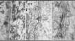
Thie top image shows clumps of intermediate filaments in the basal keratinocytes, a beautifully developed lamina lucida (electron lucent) over a dark lamina densa. The vertically oriented fine filaments are anchoring fibrils. The horizontally oriented banded structures are collagen fibrils.
The center micrograph shows the hemidesmosomal attachment plaque and the anchoring fibrils radiating downward into the dermis.
The bottom micrograph shows keratin filaments connecting to the desmosomal attachment plaque, a well developed lamina lucida, lamina densa and banded, curved anchoring fibrils.