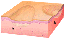


This illustration shows two examples of atrophy. In "A"there is dermal thinning without changes in the epidermis. In "B" there is thinning and flattening of the epidermis as well as dermal thinning. Epidermal atrophy allows visualization of the superficial dermal vasculature, as shown.
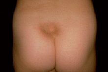
This patient has a circumscribed depressed area of skin and an otherwise unremarkable skin examination. The pigmentary change suggests that epidermal and probably dermal atrophy are present, but the great depth of the depression suggests that atrophy of the subcutaneous fat is also present. A solitary lesion such as this in a different location might suggest that the problem is a result of an injection, perhaps of corticosteroids. In this case, no injection occurred. This is an example of localized panatrophy.