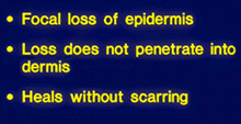
This illustration shows a partial thickness loss of the epidermis. This wound could be somewhat deeper, to include all the epidermis and still be considered an erosion. These wounds will heal without scarring, but may have pigmentary abnormalities. If the wound extends through the basement membrane zone, there will be variable amounts of scarring. An inflammatory infiltrate of cells in the dermis should accompany this lesion and could be the cause of it, or a reaction to it.
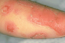
These red, shiny, erosions have a typical sunburn-like peeling desquamation at the periphery. One fresh lesion (upper left) shows that erosions often evolve from bullae. In this case, pemphigus foliaceus, due to autoantibody binding to desmoglein in the desmosomes of the granular cell layer and induction of acantholysis, is demonstrated.
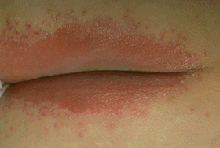
This immunosuppressed patient under treatment for cancer developed these firey red patches in an intertriginous area. The red patches are surrounded by discrete satellite papules or pustules which strongly suggests the diagnosis of candidiasis, caused by species of the yeast Candida. A Gram stain and microscopic examination of exudate from one of the pustules would show large Gram-positive budding yeast forms and an infiltrate of polymorphonuclear leukocytes.