
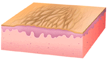
This illustration shows hyperplasia of all compartments of the epidermis with acantholysis, papillomatosis, and hyperkera tosis. It does not show thickening of the collagen bundles in the superficial dermis, and the inflammatory cell infiltrate which normally accomplanies lichenification.
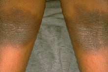
This patient has symmetrical, dark poorly defined plaques in the popliteal flexural areas. Note the grossly thickened, leathery, hyperpigmented skin and the deep, widely-spaced skin markings. This distribution of lichenification, taken with other data from the medical history and physical examination, would encourage the physician to strongly consider the diagnosis of atopic dermatitis.
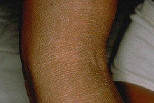
This patient has less pigmentary change than the patient illustrated in Panel C and a more widespread (extends out of the field of view) area of lichenification. In this instance, the patient had atopic dermatitis, but because of the lack of other information in this image, it is difficult to formulate a diagnostic hypothesis. The only inference that can be made is that the patient is very itchy and that he or she has been rubbing or scratching a great deal.