
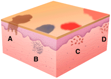
The colors of macules include shades of brown, due to melanin pigmentation in the epidermis (A), blue, due to melanin or other particulates (like some tattoo pigments) in the der mis (B), red, due to vasodilatation in the dermis without (C), and with (D) inflammatory cells present. Patches with very distinct margins tend to be due to pigmentary alterations in the epidermis, whereas those with indistinct margins tend to have a grea ter dermal inflammatory component.
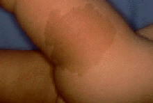
This large brown patch on the thigh and hip of a child has the uniform color of coffee with cream and thus the name of "cafe au lait" patch. This lesion is often associated with neurofibromatosis. The patch es are more darkly pigmented than the surrounding skin because there is an increase in number and activity of melanocytes in the epidermis.
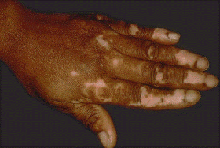
These irregular acral white patches on the fingers of a darkly-pigmented individual are completely lacking in melanocytes and are diagnostic of vitiligo, a condition in which there is autoimmune destruction of melanocytes. The faint erythema (redness) evident in the white macules and patches may be due to ultraviolet light exposure. Because of the lack of photoprotective melanocytes, the skin in these areas would be expected to be more sensitive to ultraviolet light-induced damage, sunburn, and carcinogenesis. In darkly-pigmented individuals, this is a significant cosmetic problem as well.