
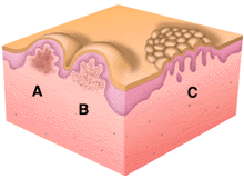
Papules are palpable because there is accumulation of material in the dermis (A and B) which gives additional substance to the skin. The accumulated material may be a metabolic deposit, amyloid or mucin, for example (A), or a cellular infiltrate of inflammatory or neoplastic cells. (B) Often the lesion's substance arises from proliferation of cells in one or more layers of the epidermis (C). Papules due to dermal metabolic deposits or cellular infiltrates tend to have indistinct margins, whereas those due primarily to epidermal hyperproliferation tend to have very well-demarcated margins. When the papule itself consists of tightly packed individual small hyperkeratotic tiny papules, the lession is said to be verrucous or warty or vegetating.
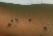
This group of variably-sized symmetrical dome-shaped and umbilicated brown papules is diagnostic of molluscum contagrisum, a viral infection that can be sexually transmitted.

These erythematous scaly papules on the abdomen are diagnostic of psoriasis, achronic inflammatory skin disease, which in this case was precipitated by streptococcal pharyngitis. These papules contain both an inflammatory dermal component and a hyperplastic epidermal component. The tendency to occur in areas of skin injury (as in the linear distribution of papules seen here, probably from a scratch) is typical for a small number of skin diseases, including psoriasis.