
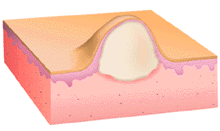
This illustration shows the same morphology as vesicle/bulla, but instead of having clear fluid within the vesicle, it is filled with pus. As with vesicles and bullae, pustules may occur at different levels within the epidermis. When the pus is contained within the dermis, the lesion is termed abscess or furuncle and when deep and dissecting through tissue planes, carbuncle.
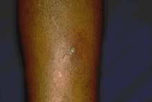
This solitary pustule is centered on a much larger area of erythema on the lower extremity. A Gram stain of the pustule contents would reveal the etiologic agent, Gram-positive Staphylococcus aureus and many polymorphonuclear leukocytes.
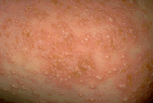
Innumerable tiny superficial, probably subcorneal, pustules occurring on an erythematous base over wide areas of skin would strongly suggest the diagnosis of generalized pustular psoriasis. A Gram stain of pus from one of these lesions would show polymorphonuclear leukocytes only, and no bacteria.
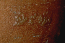
These tightly grouped tense pustules are associated with local erythema. Some of the vesicles are umbilicated. All are in the samestage of evolution. A Gram stain of pus from one of these pustules would reveal polymorphonuclear leukocytes only and no microorganisms. Except for being pustules instead of vesicles, these lesions are very similar to the vesicles of herpes simplex virus infection and are nearly diagnostic for this infection.
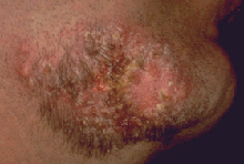
These deep-seated pustules associated with patches of alopecia (hair loss) and crusting (dried serum, see below) occurred on a discrete patch of skin in the beard area. Because the patient has not shaved in this area, one might conclude that the lesions were painful or very exudative . Though this image evokes a list of diagnostic possibilities, a deep bacterial pyoderma or an inflammatory reaction to a superficial fungal infection of the skin and hair (tinea barbae, kerion celsi) are the most likely. In this case, Gram stain of the pus showed many polymorphonuclear leukocytes and Gram-positive cocci in clusters, and did not reveal fungal organisms, leading to the diagnosis of Staphylococcal pyoderma.