
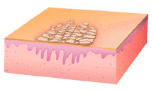
This illustration shows a plaque with overlying scale. The plaque in this case has a hyperplastic epidermis and the scales are thick, densely packed, and cracked apart, like dried mud.
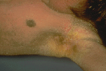
This patient has diffuse scaling with accentuation in the anterior and posterior axillary folds. Note how thick and yellow the scale is on this individual. These findings taken with a history of being chronic, and present from birth strongly suggest a diagnosis of ichthyosis.
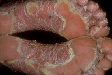
This patient has extensive scaling on the feet, with focal areas of clinically normal skin. Note that no erythema is present, but that the quality of the scale is thick and shiny and compact, in contrast to the patient in Panel C. This patient had psoriasis.