
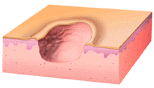
This illustration shows an ulcer. Note the complete loss of the epidermis and partial loss of the dermis. The name of the lesion doesn't change if the dermis is completely penetrated. The illustration shows a rolled-up or heaped-up border which might be the result of neoplastic growth, or inflammation and edema.
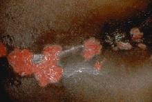
This chronically HIV-1 infected, immunoincompetent patient developed these punched out perianal ulcers with red granulating bases. The ulcer in the middle of the image has a thin greyish border that indicates early re-epithelialization of the wound. These ulcers, which look like they were made with a paper punch striking many times in nearly the same place are all in the same stage of evolution and are diagnost ic of Herpes simplex virus infection.
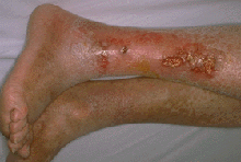
This elderly patient has many, variably sized ulcers on the lower extremity. The skin is shiny and hairless, indicating poor peripheral perfusion and has pigmentary changes suggesting that an element of venous incompetence is possibly also present (to validate that hypothesis, examine the patient with the legs in the dependent position, and look for varicosities). Some superficial areas of hemorrhage (petechiae) in the dermis are evident which buttresses the hypothesis that these ulcers have a vascular origin, and are probably due to circulatory insufficiency. A vasculitis is not easily ruled out by clinical examination, but the lack of involvement of the contralateral extremity points to the pre-eminence of local factors in the pathogenesis of these ulcers.