
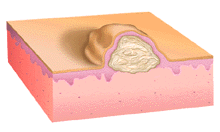
This illustration shows a fluid-filled chamber arising between the epidermis and the dermis. The fluid may be clear and straw-colored or have variable amounts of blood mixed in. Generally, vesicles arising in deeper cleavage planes are more tense and less transparent than those arising in more superficial layers of the epidermis.
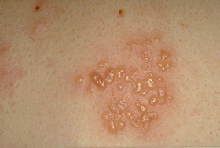
These grouped irregular tense fluid-filled vesicles appear to be all in the same stage of development. They are associated with some erythema where they have occurred. Some of the vesicles have a tiny central depression or umbilication that allows the clinical diagnosis of herpes simplex virus infection to be made with some certainty. Early, deep vesicles don't obviously contain fluid, however. Sometimes needle aspiration is required to firmly establish the vesicular nature of the lesion.
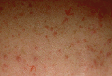
This widespread erythematous eruption is primarily papular but two distinct vesicles are also present (arrows). The distribution, size, and density of lesions taken together with the clear coexistence of both papular and vesicular elements strongly suggests the diagnosis of transient acantholytic dermatosis. Microscopic examination of stained sections of a biopsy of one of these lesions would show features of dyskeratosis (abnormal maturation of keratinocytes) and acantholysis (separation of keratinocytes by dissolution of intercellular connections). The clinical spectrum of disease is a reflection of the spectrum of pathological findings.
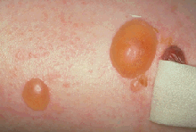
These tense symmetrical bullae contain straw-colored fluid. Another more flaccid lesion (just under the bandage) contains a small amount of blood. These bullae, unlike the ones seen in panel C, have developed on clinically normal skin. Their tenseness suggests that the origin of the vesicle is deep in the epidermis or at the junction of the epidermis and dermis. Although not diagnostic, these lesions strongly suggest the diagnosis of bullous pemphigoid and would direct a diagnostic laboratory evaluation.
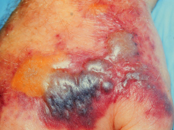
Some of these tense, subepidermal bullae are filled with straw-colored serum; others have the distinctive blue-purple color of blood. these lesions arise on an edematous, infiltrated, purpuric base. In this patient with myelodysplastic syndrome, these lesions would strongly suggest the diagnosis of acute febrile neutrophilic dermatosis (Sweet's Syndrome).