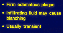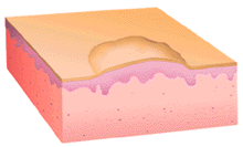


This illustration shows a raised papule that lacks the associated epidermal hyperplasia or dermal metabolic deposits or cellular infiltrates that define true papules. This lesion is due to the transudation of fluid into the epidermis due to a local, acute change in vascular permeability and is properly termed wheal.
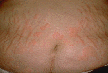
This man has annular, circular, and polycyclic wheals on his abdomen. Wheals have rather ind istinct margins, as would be expected for skin lesions where the primary pathological process is in the dermis. In this case the wheals are erythematous, indicating that vasodilatation has occurred.
These wheals are much less well-defined and show very little erythema compared to the lesions depicted in panel C. The follicular orifices appear patulous. This constellation of findings is still consistent with the diagnosis of urticaria, but suggests that the reticular dermal plexus is principally affected.