
The tissues of the skeletal system, bone and cartilage, are
specialized types of connective tissue. Unlike epithelia,
connective tissue consists of scattered cells and an abundance of
extracellular material. Connective tissue cells synthesize
and secrete proteins such as collagen that assemble into large
fibers to give structure to the tissue. Bone tissue is rigid
and strong because mineral salts of calcium are deposited within
the framework of collagen fibers. Cartilage is solid and
flexible tissue containing a large amount of proteoglycans
(sugar-linked proteins) among the collagen fibers of the
extracellular matrix. The proteoglycans contribute to the
physical properties of cartilage that allow it to resist
compression.
At the gross level, it is possible to distinguish two basic types
of bone tissue: compact bone
and trabecular
bone (trabecular bone is also called
spongy bone or cancellous bone). Compact bone is dense and
solid, while trabecular bone consists of a fine network of
interlocking struts. Compact bone forms the outer layer of
long bones, while trabecular bone is found in the central cavity,
and also at the ends of bones. The figure below shows a
tibia that has been sectioned longitudinally, so that you can
compare compact bone to trabecular bone.
There are three basic types of cells found in bone: osteoblasts, osteocytes, and osteoclasts (see page about bone remodeling). The slides below are of thin ground sections of dried bone, and so the living cells are not visible. However, this type of preparation is good for revealing the spaces and fine channels that are occupied by osteocytes (bone cells) in the living bone.
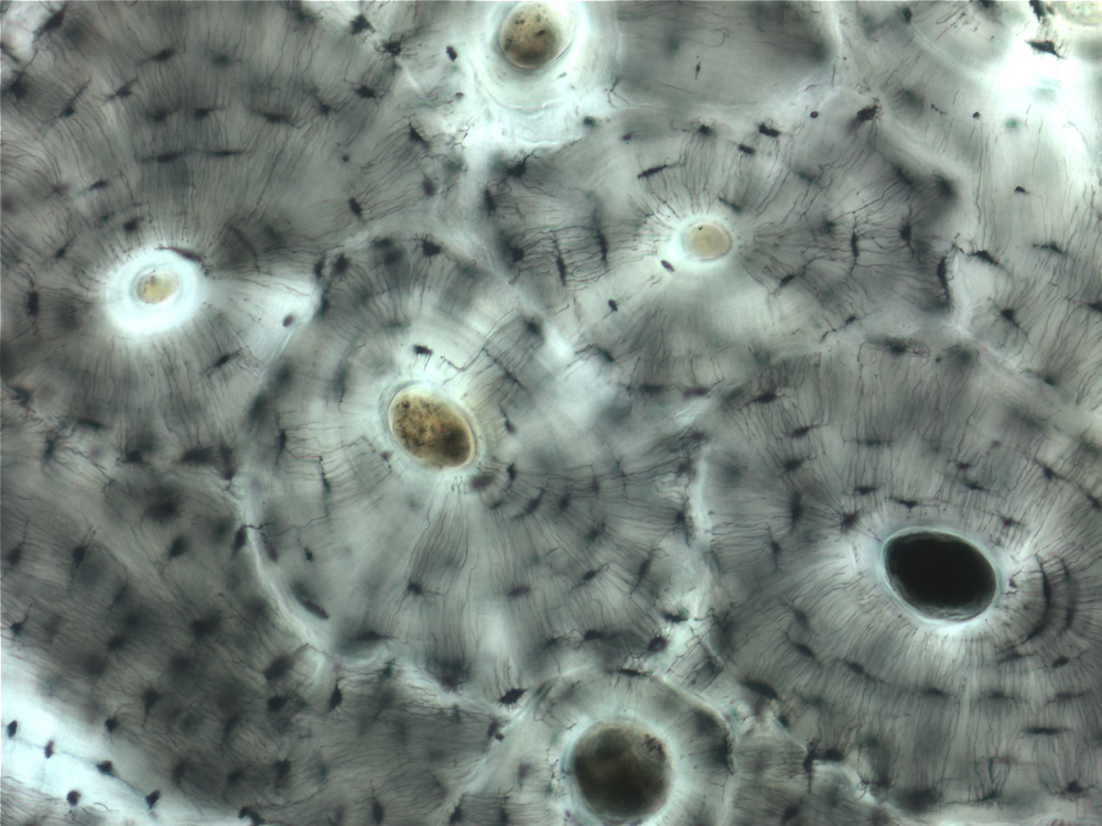 This picture shows
compact bone at low magnification. Compact bone consists of
many parallel units called osteons
that are tightly packed together to form the strong outer layer of
bone. The large circles that you see at the centers of the
osteons are channels known as Haversian
canals. In the living tissue, the Haversian
canals are occupied by blood vessels and nerves. Each osteon
consists of concentric layers of bone tissue surrounding a
Haversian canal. For a schematic view showing the
organization of bone tissue, look at Figure 10.10 in Wheater's
Functional Histology (see lecture slides).
This picture shows
compact bone at low magnification. Compact bone consists of
many parallel units called osteons
that are tightly packed together to form the strong outer layer of
bone. The large circles that you see at the centers of the
osteons are channels known as Haversian
canals. In the living tissue, the Haversian
canals are occupied by blood vessels and nerves. Each osteon
consists of concentric layers of bone tissue surrounding a
Haversian canal. For a schematic view showing the
organization of bone tissue, look at Figure 10.10 in Wheater's
Functional Histology (see lecture slides).
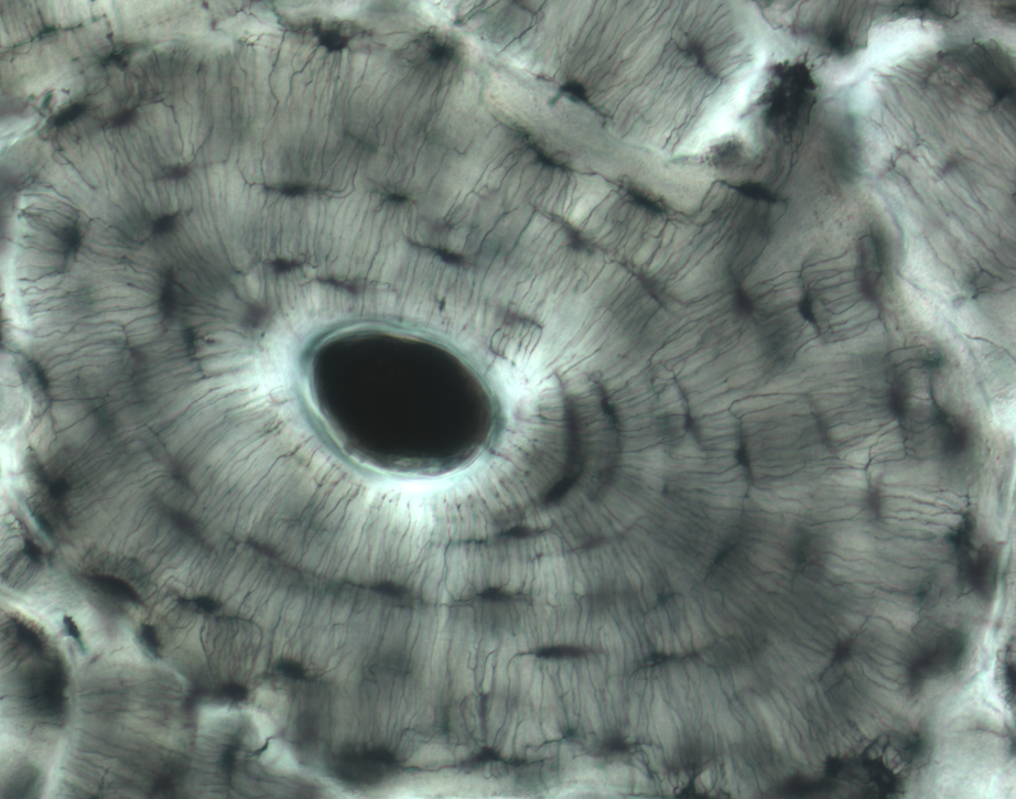 In the higher
magnification view at right, you can see that there are black
spaces arrayed around the central Haversian canal. These
spaces are called lacunae
(singular: lacuna)
and contain osteocytes in
living bone. Because osteocytes are enclosed within layers of
bone, they need to have a way to receive nutrients and communicate
with other cells. So each osteocyte extends fine cellular
processes through narrow channels called canaliculi
(singular: canaliculus).
In the slide at right, the canaliculi are the fine black lines
extending perpendicularly from each lacuna.
In the higher
magnification view at right, you can see that there are black
spaces arrayed around the central Haversian canal. These
spaces are called lacunae
(singular: lacuna)
and contain osteocytes in
living bone. Because osteocytes are enclosed within layers of
bone, they need to have a way to receive nutrients and communicate
with other cells. So each osteocyte extends fine cellular
processes through narrow channels called canaliculi
(singular: canaliculus).
In the slide at right, the canaliculi are the fine black lines
extending perpendicularly from each lacuna.
Cartilage is found in a layer at the ends of bones where they form joints (the articular cartilage; we will look at joints next week). Cartilage also forms structural rings and plates in the large airways of the respiratory tract. The slides below are sections from the trachea.
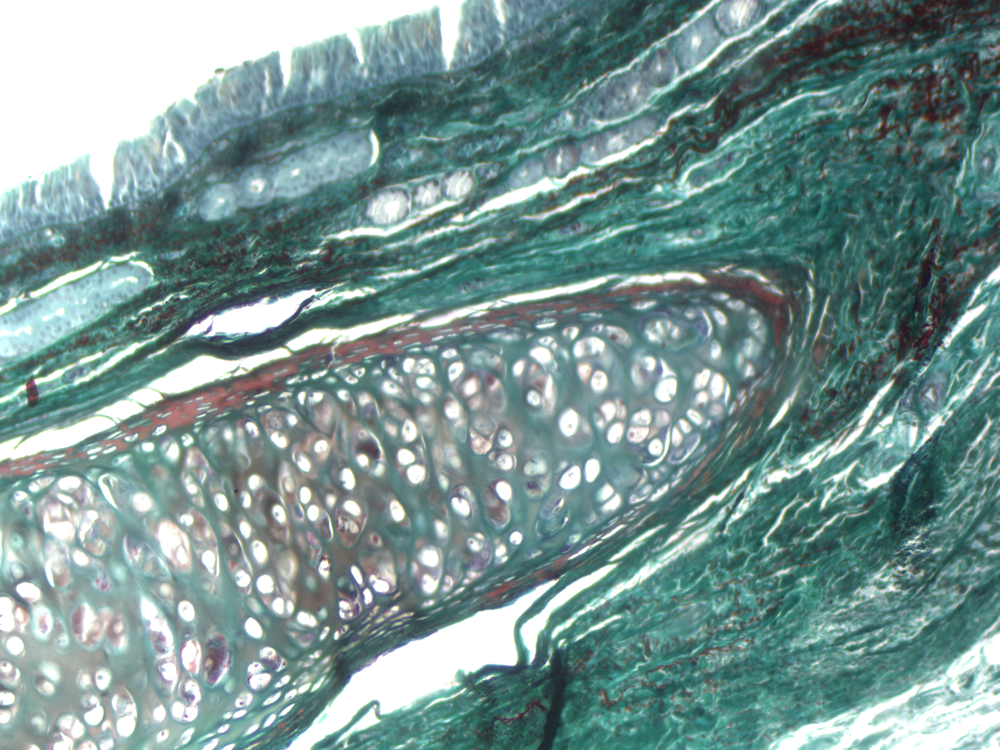 The trachea is the large
central airway that connects the upper respiratory tract (nasal
passages, oral cavity, throat structures) to the airways that lead
to the lungs. The trachea is held open by rings of cartilage.
These rings do not form a complete circle; instead they have the
shape of a C. The picture at right shows the end of one of the
C-shaped rings of cartilage extending off to the lower left.
The trachea is the large
central airway that connects the upper respiratory tract (nasal
passages, oral cavity, throat structures) to the airways that lead
to the lungs. The trachea is held open by rings of cartilage.
These rings do not form a complete circle; instead they have the
shape of a C. The picture at right shows the end of one of the
C-shaped rings of cartilage extending off to the lower left.
The specific type of cartilage found in the trachea and in the
articular cartilage is called hyaline
cartilage. Hyaline means glassy, and this
type of cartilage has a smooth and translucent appearance.
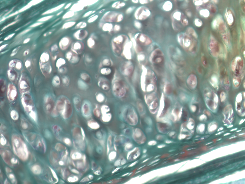 At the microscopic level,
cartilage has the appearance of Swiss cheese. In the living
tissue, each space that you see is occupied by a cartilage cell,
called a chondrocyte. As in
bone tissue, the spaces are called lacunae
(singular: lacuna).
Unlike bone, cartilage is not penetrated by blood vessels. This
limits how thick cartilage can become since nutrients must diffuse
through the tissue to reach cells. Cartilage also has much less
capacity for repair. Damage to articular cartilage is what
occurs in arthritis.
At the microscopic level,
cartilage has the appearance of Swiss cheese. In the living
tissue, each space that you see is occupied by a cartilage cell,
called a chondrocyte. As in
bone tissue, the spaces are called lacunae
(singular: lacuna).
Unlike bone, cartilage is not penetrated by blood vessels. This
limits how thick cartilage can become since nutrients must diffuse
through the tissue to reach cells. Cartilage also has much less
capacity for repair. Damage to articular cartilage is what
occurs in arthritis.