Epithelial Histology
An epithelium is the type of tissue that covers surfaces,
usually the linings of hollow organs in the body, or in the case
of the skin, the outer surface of the body. In many cases,
adjacent epithelial cells are linked by tight
junctions so that the epithelium forms a barrier
that regulates the movement or substances across it (see the web
page on Epithelial
Transport). The figure below shows a schematic of a
simple epithelium, modeled after the epithelium lining the small
intestine.
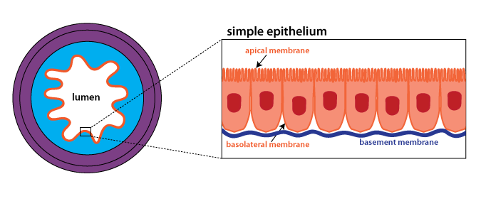
Epithelia are comprised almost entirely of cells. The apical surface faces the lumen (inside of a hollow organ)
while the basal or basolateral surface is adjacent to
the underlying tissue. In many epithelia, the apical surface
is specialized. For instance, in the small intestine, the
apical plasma membrane is folded into microvilli
(see below) to increase the surface area for absorption of
nutrients. Every epithelium has a basement
membrane, that binds the epithelium to the
underlying connective tissue. The basement membrane consists
of a thin layer of extracellular proteins that is located next to
the basolateral surface. Blood vessels do not penetrate
through the basement membrane. In practice, the basement
membrane is too small to be seen except on an electron micrograph
(EM). In the lecture we will look at an EM of the skin
epithelium that shows the basement membrane.
Epithelia are classified according to whether they consist of a
single layer of cells (simple) or multiple layers of cells
(stratified). Epithelia are also classified by the shape of
the cells: squamous (flat), cuboidal, or columnar.
Be able to identify the following examples, where they are found
in the body, and what type of epithelium they are
(for example: simple squamous epithelium). Terms shown
in purple boldface may appear
on a quiz section test.
In the textbook*, figure 3.10 (p. 78) is a useful overview of
different types of epithelia. In this figure, epithelia are
classified according to function, as opposed to shape and cell
number. Figure 3.15 (p. 86) illustrates the structure of the skin.
*Pages and figure numbers refer to the 8th
edition of Human Physiology: An Integrated Approach by
Dee Silverthorn.
Examples
Epidermis of the Skin

The figure at right shows a low-magnification view of thick skin,
such as you would find on the surface of your fingertip where your
fingerprint is. At this low magnification you can see the
three layers in the skin. The outer region is the epidermis, which forms the surface
of the skin. The middle region is the dermis, which contains
dense connective tissue, blood vessels, and nerves. The
innermost layer is the hypodermis, which contains loose connective
tissue and adipose tissue, along with sweat glands, blood vessels,
and nerves.
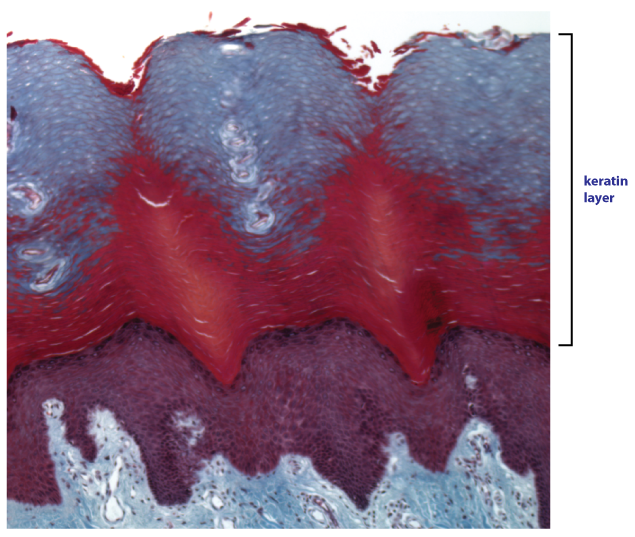
This next image is at higher magnifcation and focuses in on the
epidermis. The epidermis is classified as a stratified
squamous keratinized epithelium. Cells divide
at the basal surface of the epithelium, and as new cells are
generated, older cells are pushed towards the apical
surface. As cells move apically, they become very flattened
(squamous) and they fill up with an intracellular protein called keratin (for this reason, cells in
the epidermis are referred to as keratinocytes).
This
type
of epithelium is protective, with the keratin
layer acting to protect the underlying tissue from
abrasion and dehydration.
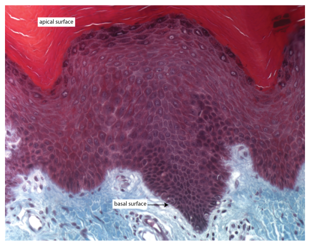
This image focuses on the keratinocytes
(purple layer) at high magnification. Note that cells appear
dark at the apical surface, where they are filling up with
keratin.
OPTIONAL: Melanocytes, which produce pigment, are
located on the basal surface of the epidermis. Although they
produce pigment, their cytoplasm appears clear in this stain. See
if you can find a melanocyte in this picture.
Endothelium
The endothelium is the simple squamous epithelium that
lines all blood vessels. The very smallest vessels,
capillaries, are made up of just endothelium. A healthy and
intact endothelium acts as a barrier that isolates the blood from
underlying tissue, preventing the formation of a blood
clots. Endothelial cells also send important signals,
affecting things such as tissue growth and blood flow. In
the brain, there are tight junctions
between endothelial cells, limiting the permeability of the
endothelium, and protecting neurons in the brain from toxins and
other potentially neuroactive substances in the blood. This
functional barrier is known as the blood-brain
barrier.
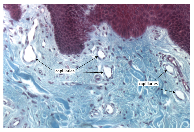
The image below shows capillaries in the upper part of the
dermis. Note how each white space is outlined by a single
layer of thin dark cells; this is the endothelium.
Small Intestine
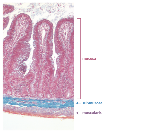 This low-magnification view shows the different
regions of the small intestine. In the digestive tract and
other organs, the layer of tissue next to the lumen is called the
mucosa. The mucosa is sometimes called a "mucous
membrane" because it is covered with a layer of mucus. The
mucosa consists of a small amount of connective tissue and a
highly folded simple columnar epithelium.
The folds of tissue that poke out are called villi
(singlular: villus), and the folds that dip down are called
crypts. The folding of the epithelium increases the surface
area available for absorption of nutrients.
This low-magnification view shows the different
regions of the small intestine. In the digestive tract and
other organs, the layer of tissue next to the lumen is called the
mucosa. The mucosa is sometimes called a "mucous
membrane" because it is covered with a layer of mucus. The
mucosa consists of a small amount of connective tissue and a
highly folded simple columnar epithelium.
The folds of tissue that poke out are called villi
(singlular: villus), and the folds that dip down are called
crypts. The folding of the epithelium increases the surface
area available for absorption of nutrients.
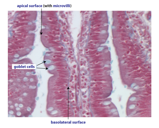
The image at the left shows part of a villus at high
magnification. The absorptive cells (pinkish red) in the simple columnar epithelium are
called enterocytes.
Scattered among the enterocytes are mucus-secreting cells called goblet cells that stain pale
blue. At the apical surface of the enterocytes, there is a
dark line which occurs due to a web of proteins that anchor the microvilli, folds of the apical
plasma membrane that further increase the surface area for
absorption. Another term for the microvilli is "brush
border". We will look at several EMs that show microvilli in
the lecture.
Airway Epithelium
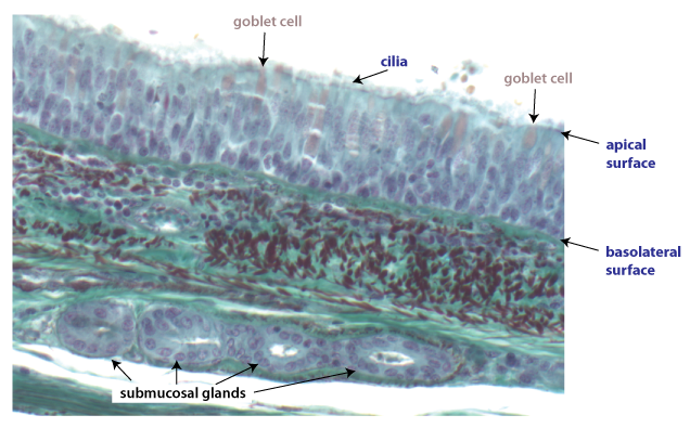 The airway epithelium
lines the passages that conduct air into the lungs. (Airways
conduct air into the lungs whereas tiny sacs called alveoli are
where gas exchange occurs.) The airway epithelium is
classified as a pseudostratified
ciliated epithelium. Although it is a simple
epithelium, it appears stratified because nuclei of different
cells are at different heights, hence the term
"pseudostratified". An important function of this epithelium
is the movement of mucus out of the respiratory tract.
Mucus, which is secreted by goblet cells and submucosal glands,
traps particles and pathogens. The beating of the cilia, located at the apical surface, moves the mucus
toward the pharynx (throat) where it can be swallowed.
The airway epithelium
lines the passages that conduct air into the lungs. (Airways
conduct air into the lungs whereas tiny sacs called alveoli are
where gas exchange occurs.) The airway epithelium is
classified as a pseudostratified
ciliated epithelium. Although it is a simple
epithelium, it appears stratified because nuclei of different
cells are at different heights, hence the term
"pseudostratified". An important function of this epithelium
is the movement of mucus out of the respiratory tract.
Mucus, which is secreted by goblet cells and submucosal glands,
traps particles and pathogens. The beating of the cilia, located at the apical surface, moves the mucus
toward the pharynx (throat) where it can be swallowed.