Anatomy of the Urinary System
The goal this week is to learn the gross anatomy of the kidney
and urinary tract. In lecture, we will look at the histology
of the nephron, which is the functional unit of the
kidney.
Recommended Reading
Read section 19.1 and 19.2 pp. 588-592 in Silverthorn (function
of the kidneys and anatomy of the urinary system).
Gross Anatomy of the Kidney
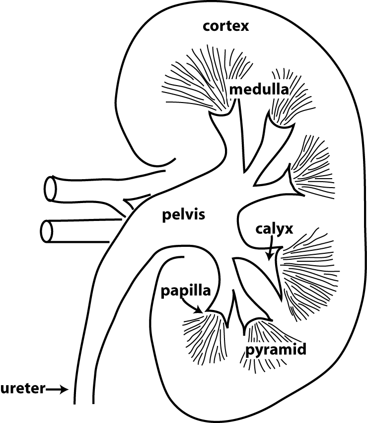
The figure at the right depicts the structures that are visible
when you section a kidney along its long axis. There are two basic
regions in the kidney, an outer cortex,
and an inner medulla.
As you will see, certain parts of the nephrons are located in the
cortex, and certain parts are located in the medulla.
The medulla contains dark structures known as pyramids.
Each pyramid has a tip known as the papilla.
Urine forms in the nephrons and drains from collecting ducts in
the papilla. The papilla fits into a cup-like structure called a calyx. The calyces form
short branches that attach to a wide space called the renal pelvis. The renal
pelvis then narrows to form the ureter,
a tube that conveys urine to the bladder
where it is stored. The calyces, renal pelvis, ureters, and
bladder are all lined with a specialized type of epithelium called
uroepithelium (or transitional epithelium).
The uroepithelium is a stratified epithelium with tight junctions
between adjacent cells so that it acts as an important
permeability barrier. Urine can be made more
concentrated than the extracellular fluid, so it is
important that the structures that conduct and store urine don't
allow water to be drawn into the urine. Another unique
feature about the uroepithelium is that the epithelium can stretch
when the bladder is full and its walls are distended. In
class we will look at the uroepithelium when we review kidney
histology.
The key function of the kidneys is to regulate the composition
and volume of the extracellular fluid. Critical to this function
is a high rate of blood flow. The renal blood flow is typically
about a fifth of the total cardiac output. The renal artery carries blood
from the aorta to the kidney, and the renal
vein returns blood from the kidney to the inferior
vena cava and heart. The renal artery, renal vein, and renal
pelvis are all located in a region of the kidney called the hilum.
Model of the Kidney
Be able to identify the following structures in the model of the
kidney:
In the model of the kidney find:
cortex
medulla
papilla
calyx
renal pelvis
ureter
renal artery
renal vein
Optional (but helpful): Kidney Anatomy Video in Acland's
Video Atlas of Anatomy
The following video is helpful for understanding kidney
anatomy. It is optional because I won't be testing you
specifically on content from the video. The link should
open the video in a new tab.
"Kidneys"(4:44) Video 5.2.27
Model of the Urinary Tract
The second model shows the the major blood vessels, and the
urinary tract. The ureters
connect the kidneys to the bladder,
which stores urine. The urethra
is the tube that carries urine from the bladder to the outside of
the body. In males, the urethra passes through the prostate
gland and penis, and also conveys semen. What is the gender
of the model?
The bladder is sectioned to show you the structures in its
interior. The wall of the bladder consists of the detrusor muscle, which is smooth
muscle, with an overlying mucosal layer that has
uroepithelium on its surface. The trigone
is a thickened region of bladder wall on the posterior side near
the base of the bladder. The openings
of each ureter are in the lateral portion of the
trigone. The ureters penetrate the wall of the bladder in an
oblique orientation, and their openings are narrow and
slit-like. This arrangement ensures that the openings of the
ureters will squeeze shut when the detrusor muscle contracts
during urination. This prevents reflux of urine back towards
the kidneys.
kidney
renal artery
renal vein
inferior vena cava
aorta
ureter
bladder
detrusor muscle
trigone
opening of ureter (in trigone)
prostate gland
prostatic urethra
Urinary Anatomy: Clinical Example
The first segment of the urethra in males passes through the prostate gland and is called the prostatic urethra (refer to figure
26.7b on p. 812 of the textbook). As men age, many will
experience non-cancerous growth of the prostate gland called benign prostatic hyperplasia.
Benign prostatic hyperplasia can cause difficulty in urination
because the enlarged prostate presses on the urethra. Common
symptoms are increased frequency of urination, difficulty in
starting urination or fully emptying the bladder, and a weak flow
of urine.
The prostate gland is in a difficult place to access surgically,
so benign prostatic hyperplasia is usually treated with drugs.
- The initial approach to treatment is to promote relaxation of
smooth muscle surrounding the bladder outlet and urethra. Smooth
muscle in the urethra and prostate gland is innervated by sympathetic neurons, which
excite smooth muscle contraction. Thus, alpha-adrenergic
antagonists that block this excitation are used
to relax smooth muscle and promote urine flow.
- In some cases, the prostate gland volume is much increased, so
treatment that limits further prostate growth is desirable. The
prostate gland grows in response to a type of androgen hormone
known as dihydrotestosterone
(DHT). 5-alpha-reductase inhibitors are
enzyme inhibitors that reduce the formation of DHT in order to
limit prostate growth.