Cells in the Nervous System
Recommended Reading
Read section 8.2, Cells of the Nervous System (pp. 226-233) in
Silverthorn. Focus on figure 8.5a (p. 230) to learn about the
different types of glial cells.
Neurons
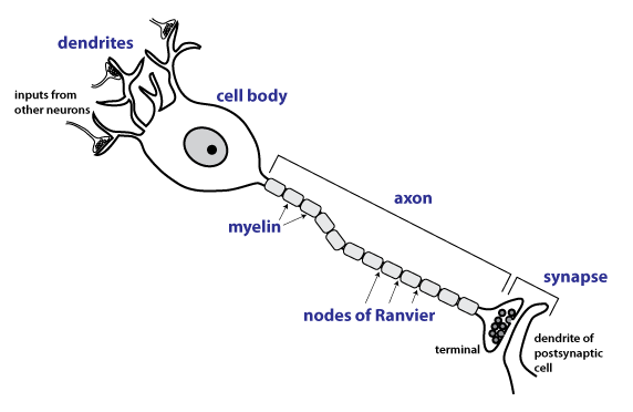
The figure below shows the parts of a typical neuron. Be
able to identify the terms in purple
boldface in the figures below.
- The dendrites of a neuron
constitute an input region where a neuron receives synapses from
other neurons. Afferent neurons that sense touch,
temperature and pain in the skin have sensory dendrites
containing specialized ion channels that open in response to the
particular sensory modality. For instance the sensory
dendrites of touch-sensitive neurons have mechanically-gated ion
channels.
- The cell body is the
metabolic center of the neuron. In general neurons are
large cells and some have axons that are very long, so
therefore, neurons have large cell bodies. Neurons are
often actively synthesizing lots of protein so they have an
extensive network of rough endoplasmic
reticulum (rough ER)
and a large and prominent nucleolus
(the organelle inside the nucleus where ribosomal RNA is made).
- The axon is part of the
output region of the neuron. The axon conducts
action potentials and thus contains voltage-gated
ion channels in its plasma membrane. Some axons can be up
to a meter long.
- Many axons are myelinated. Myelination
speeds axonal conduction. Myelin
consists of many tightly wrapped layers of plasma membranes (see
the electron micrographs shown in the lecture). Myelin
forms in bundles covering short segments of the axon; the space
between two bundles of myelin is called a node
of Ranvier.
- At the axon terminal, there is a synapse
with a postsynaptic neuron or other target cell. In an
electron micrograph, presynaptic terminals can be identified by
the presence of synaptic vesicles. Neurons may branch near
their terminals and form synapses onto multiple postsynaptic
cells.
The Histology Guide: Virtual Histology Guide
has an excellent slide
of the spinal cord that we will look at in
class. We will focus on the large somatic
motor neuron cell bodies found in the ventral horn of the
spinal cord. This tissue in this slide is stained with a
basic dye that stains
the rough endoplasmic reticulum and the nucleolus.
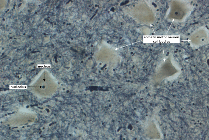 At right is high magnification view of somatic
motor neuron cell bodies in the spinal cord. This tissue is
stained by a method that produces a dark black stain on the
myelin. The neuronal cell bodies are the pale, tan
structures, and the nucleus can be made out as a slightly lighter
region within the cell body. Inside the nucleus is the dark
black nucleolus, an organelle
where ribosomes are made.
At right is high magnification view of somatic
motor neuron cell bodies in the spinal cord. This tissue is
stained by a method that produces a dark black stain on the
myelin. The neuronal cell bodies are the pale, tan
structures, and the nucleus can be made out as a slightly lighter
region within the cell body. Inside the nucleus is the dark
black nucleolus, an organelle
where ribosomes are made.
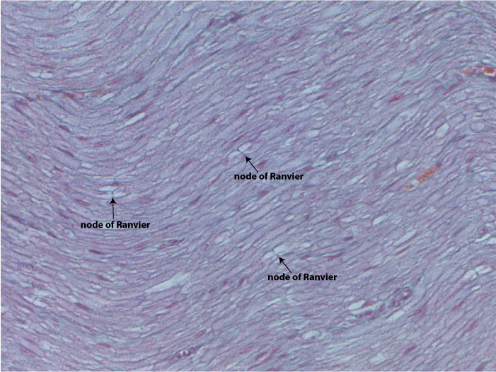
The picture below is high magnification of a longitudinal section
from a peripheral nerve. The axons are running across the view
from right to left. During histological processing, the
lipid is mostly dissolved, so the bundles of myelin look like
empty bubbles. When the section passes through a node of
Ranvier, it will appear as a line running perpendicular to the
axon.