CNS Anatomy: Spinal Cord
Overview
The goal in this exercise is to learn the anatomy of the spinal
cord, and associated nerves and roots. The function of these
different parts will be laid out in the web page about the
Organization of the Peripheral Nervous System.
Recommended Reading
Read section 9.4 pp. 281-282 in Silverthorn (general organization
of the spinal cord).
Spinal Cord Tissue
The most important terms of direction for studying spinal cord
anatomy are ventral (which means "towards the stomach")
and dorsal (which means "towards the back"). Note
that in human anatomy, the ventral side of the spinal cord
corresponds to the anterior side, and the dorsal side corresponds
to the posterior side.
The tissue in the spinal cord, like the brain, can be divided
into gray matter, containing
primarily neuronal cell bodies, and white
matter containing axons, arranged into tracts.
The
gray
matter is located centrally and has roughly the shape of a
butterfly. The white matter is around the outside. The central canal is a space
containing cerebrospinal fluid that links to the ventricles in the
brain.
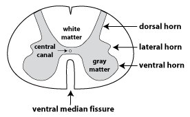
The figure at the right depicts a cross-section from the thoracic
region of the spinal cord. The ventral side can be identified by
looking for the ventral median fissure.
As
well,
the ventral horn of the gray
matter is rounded, while the dorsal horn
is usually narrower and extends out to the edge of the spinal
cord. The lateral horn, which
is found mainly in the thoracic region of the spinal cord,
contains the cell bodies of autonomic efferent neurons,
specifically sympathetic preganglionic
neurons.
The shape of the gray matter varies in different regions of the
spinal cord. The figures below are low magnification views of
histology slides of the spinal cord. The tissue has been treated
with a black stain specific to myelin, so that the white matter
appears dark, and the gray matter appears light.
Thoracic Spinal Cord
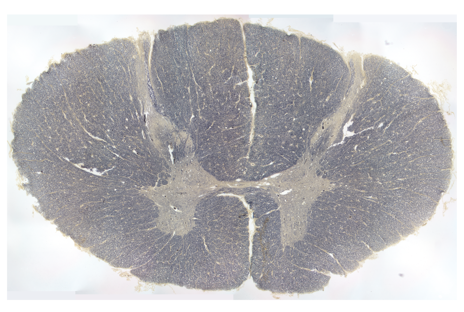
This section is from the thoracic region. This figure and the one
below are oriented so that dorsal is at the top, and ventral is at
the bottom. The dorsal horn
is narrow and extends out to the edge of the spinal cord, while
the ventral horn is
rounded. There is also a lateral
horn at this level of the spinal cord.
Sacral Spinal Cord
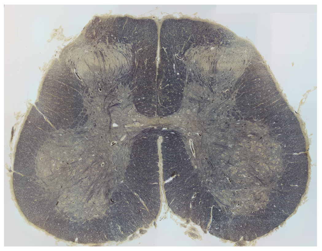
This section is from the sacral region. This part of the spinal
cord contains efferent neurons that control the muscles of the
legs, and so has many more somatic motor neurons in the ventral
horn. Likewise, there is much more afferent input from the legs
than from the trunk. Consequently, both the dorsal
horn and ventral horn
are much larger than in the thoracic segment. The ventral median fissure can be seen
in this section, but is harder to see in the thoracic section
above.
Models of the Spinal Cord
There are two spinal cord models that we will study. When not
being used in our class this week, these models are available on
the third floor of the Health Sciences Library. Pictures of
the models are provided in the links here, and also in the lecture
slides.
The first model depicts a cross-section of the spinal cord at the
cervical level (in the neck). The spinal cord sits within the vertebral
canal which is the passageway within the bones (vertebrae)
of the spinal column. This model depicts the three layers of
the meninges (dura mater, arachnoid mater and pia mater). We will
only identify the tough, thick outermost layer, known as the dura mater. The dura mater
encloses a layer of cerebrospinal fluid (CSF) that cushions the
brain and spinal cord. This space containing CSF is called the subarachnoid space because the
arachnoid layer is attached just underneath the dura mater (see
video 3.1.8 below). In the head, the dura mater is attached
to the skull bones, but in the spinal canal, there is an epidural space filled with fat and
blood vessels that lies between the dura mater and the vertebral
bones.
The dorsal root ganglion is
the swelling that is found along the dorsal root. The dorsal
root ganglion contains the cell bodies of afferent neurons
(see the next page on the organization of the peripheral nervous
system). The dorsal root ganglia are located just outside
the dura matter. Note that the dorsal root ganglion does not
lie in a position that is dorsal to the spinal cord, but rather
gets its name because it is a cluster of neuronal cell bodies
("ganglion") that surrounds the dorsal root. Distal to the dorsal
root ganglion, the two roots fuse to form a spinal
nerve.
white matter
gray matter
dorsal horn
ventral horn
central canal
ventral median fissure
dorsal root
ventral root
dorsal root ganglion
spinal nerve
dura mater
subarachnoid space
epidural space
The second model is a model of a longitudinal dissection of the
spinal cord. All of the tissues ventral to the spinal cord are
removed, thus, we are looking at the ventral surface of the spinal
cord. On one side of the model, the roots are cut, revealing the dorsal roots; the ventral roots are visible on the
surface on the undissected side.
Click here
to see a full view of this model.
The dorsal and ventral roots travel some distance caudally before
they join to form a spinal nerve that exits the vertebral canal.
These distances become longer for more caudal segments. The tissue
of the spinal cord only extends to the beginning of the lumbar
region, so the most caudal part of the vertebral canal is a group
of long roots known as the cauda equina
(because it resembles a horse's tail). A white wavy line
represents the dura mater on this model; thus the space adjacent
to the spinal cord is the subarachnoid space. A spinal tap
is when a needle is inserted into the subarachnoid space in order
to take a sample of the CSF. This is done in the region of
the cauda equina in order to reduce the risk of damage to the
spinal cord.
The dorsal root ganglia are small, subtle, and difficult
to identify in these pictures or the model. However, the spinal nerves are easy to see as
the many white structures extending laterally away from the spinal
column. There are larger numbers of axons in the spinal
nerves leaving from spinal segments that supply nerves that
innervate the arms and the legs. In these regions, the
spinal nerves divide and rejoin to form a network called a plexus.
The
more superior plexus whose nerves innervate the arms is called the
brachial plexus; the lower one whose nerves innervate the
legs is called the lumbosacral plexus.
On the model's right side, you can see the sympathetic chain ganglia (there
are sympathetic chain ganglia on both sides of the body, but they
are only modeled on the right side for this particular model).
These ganglia contain the cell bodies of sympathetic
postganglionic neurons. In the model the
sympathetic chain ganglia are bright pink and connected by yellow
processes.
In the thoracic
region of the longitudinal dissection of the spinal cord
find:
ventral root
dorsal root
spinal nerve
subarachnoid space
sympathetic chain
ganglion (location of cell bodies sympathetic
postganglionic neurons)
In the caudal
region of the longitudinal dissection of the spinal cord
find:
cauda equina
spinal nerve
subarachnoid space
sympathetic chain ganglion
Videos from Acland's Video Atlas of Human Anatomy
The Acland's Video Atlas of Anatomy provides narrated video
demonstrations using high quality cadaver dissections. These
videos contain more detail and terminology than I expect you to
learn for this class. In each case, focus on being able to
identify the terms given in blue boldface in the yellow
boxes. Dr. Acland speaks with a British accent, so you may
want to utilize the closed captions. A pdf transcript is
also available for each video.
The first video (video 3.1.8) shows a cross section of the spinal
column. This video shows very nicely the relationship of the three
layers of meninges.
What to identify in video 3.1.8 (opens in a new
tab):
spinal cord
dura mater
subarachnoid space
epidural space
The second video (video 3.1.9) is a posterior (dorsal) view of
the spinal cord. This video shows the end of the spinal cord
tissue and the cauda equina. It also shows how the
"filaments" (nerve rootlets) leaving the dorsal and ventral sides
of the spinal cord coalesce to form the dorsal and ventral roots,
and then how the roots come together to form a spinal nerve.
What to identify in video 3.1.9 (opens in a new
tab):
spinal cord
cauda equina
dorsal root
ventral root
spinal nerve
dorsal root ganglion
Tips
I think the best views of the dorsal and ventral roots can be
seen in the section "Dura around emerging spinal nerve". To put
this in the best context, jump to the previous section "Spinal
nerves: emergence from intervertebral foramen. Note: the
intervertebral foramen is the passageway formed at the side
when two vertebrae come together. This passageway contains
the nerve roots and the dorsal root ganglion. Also note that
the Acland's videos are looking at the spinal cord from the dorsal
side, whereas the longitudinal dissection of the spinal cord model
exposes the ventral surface of the spinal cord.
Video 3.1.10 (opens in a new
tab) provides a review. Remember you will only be tested on the
structures listed above.