Anatomy of the Heart
The pictures below are from dissections of fresh pig
hearts. Hopefully these will allow you to appreciate the
nature of the tissues in the heart much more effectively than you
ever could by just looking at a model.
Recommended Reading
Read pages 440-446 in Silverthorn, focussing on figure 14.5.
External Features of the Heart
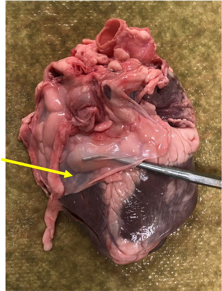 The heart is contained within a thin
membranous sac, the pericardium.
The small picture at right shows a bit of the cut pericardium on
the posterior surface of the heart. Optional: To
get
a sense of the relationship of the heart and the pericardium in
the body, you can look at Video 5.1.9 from Acland's
Video Atlas of Anatomy (the link opens in a new tab).
The heart is contained within a thin
membranous sac, the pericardium.
The small picture at right shows a bit of the cut pericardium on
the posterior surface of the heart. Optional: To
get
a sense of the relationship of the heart and the pericardium in
the body, you can look at Video 5.1.9 from Acland's
Video Atlas of Anatomy (the link opens in a new tab).
The picture below shows an anterior view of the heart with the
pericardium removed. The letters indicated in the text refer
to the labels on the picture.
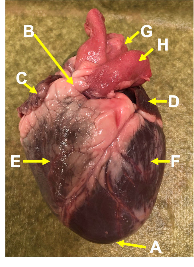 The
anterior surface of the heart is characterized by the presence of
the large arteries leaving the base of the heart, the pulmonary trunk (H) and the aorta (G). The pulmonary
trunk is the vessel that divides to give rise to the two pulmonary
arteries going to each lung. The pulmonary trunk is somewhat
anterior to the aorta, and although it is connected to the right
ventricle, it tilts leftward. The aorta is slightly
posterior to the pulmonary trunk and bound to it by connective
tissue. Note that when you look at the anterior surface of
the heart (or at anatomical illustrations of the heart in books or
on the internet) the right side of the heart will be on your left,
and the left side of the heart will be on your right.
The
anterior surface of the heart is characterized by the presence of
the large arteries leaving the base of the heart, the pulmonary trunk (H) and the aorta (G). The pulmonary
trunk is the vessel that divides to give rise to the two pulmonary
arteries going to each lung. The pulmonary trunk is somewhat
anterior to the aorta, and although it is connected to the right
ventricle, it tilts leftward. The aorta is slightly
posterior to the pulmonary trunk and bound to it by connective
tissue. Note that when you look at the anterior surface of
the heart (or at anatomical illustrations of the heart in books or
on the internet) the right side of the heart will be on your left,
and the left side of the heart will be on your right.
The inferior part of the heart is called the apex (A) because it comes to a
point, like the apex of a cone. The superior part of the heart is
referred to as the base (B).
The major vessels of the heart are found at the base of the heart,
along with the upper chambers, the right
atrium (C) and left
atrium (D). The atria are collapsed, but in a
functioning heart, they would be stretched full of blood.
The majority of the heart tissue consists of the
ventricles. The left ventricle
(F) is stiff and solid because it is very thick-walled. By
contrast, the right ventricle
(E) has thinner walls, and would collapse a little if you poked
it.
Internal Features of the Heart
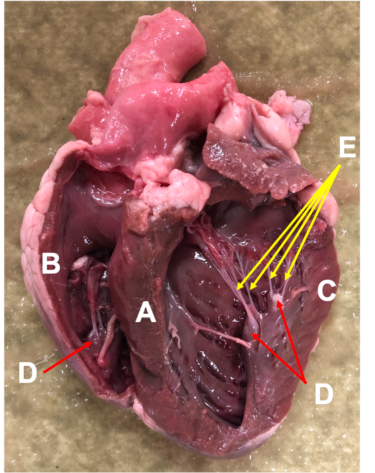 The figure at
right shows a sectioned heart where you can see the internal
structures of the heart. The left and right ventricles are
separated by the large interventricular
septum (A). Note the relative thickness of
the walls of the right ventricle (B) and left
ventricle (C). The inner surface of the
heart is covered with irregular bands of tissue known as
trabeculae carneae. The papillary
muscles (D; red arrows) are distinct
little hills of muscle that poke up from the inner surface of the
heart. At the tips of the papillary muscles are the chordae tendineae (E;
yellow
arrows), strings of connective tissue that
attach to the edges of the atrioventricular valves (AV valves).
The figure at
right shows a sectioned heart where you can see the internal
structures of the heart. The left and right ventricles are
separated by the large interventricular
septum (A). Note the relative thickness of
the walls of the right ventricle (B) and left
ventricle (C). The inner surface of the
heart is covered with irregular bands of tissue known as
trabeculae carneae. The papillary
muscles (D; red arrows) are distinct
little hills of muscle that poke up from the inner surface of the
heart. At the tips of the papillary muscles are the chordae tendineae (E;
yellow
arrows), strings of connective tissue that
attach to the edges of the atrioventricular valves (AV valves).
The heart contains four important valves that ensure that
blood flows in one direction. The right
AV valve (tricuspid valve)
and the left AV valve (mitral valve) lie between the atria
and ventricles, and prevent blood from flowing back into the atria
when the ventricles contract (systole).
The pulmonary valve and the aortic valve lie at the beginning
of the large outflow vessels at the base of the heart, and prevent
blood from falling back into the heart when the ventricles are
relaxed (diastole).
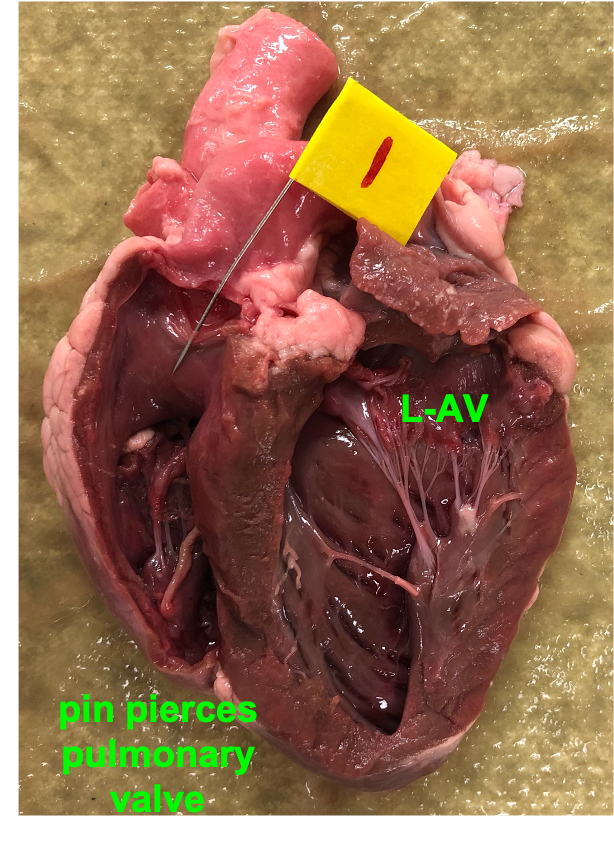 The valves consist of two
or three cusps (also called leaflets) of delicate connective
tissue that come together to block the flow of blood. The
valves in the vessels (called the semilunar valves) each have
three valve cusps that look like little pockets or like half-moons
(hence the name "semilunar"). The valve cusps of the AV
valves look like torn curtains, and are easily identified because
they have the chordae tendineae attached to them. All four
heart valves are located at the same plane in the heart and are
attached to a fibrous layer of connective tissue called the cardiac
skeleton. This is shown in the illustration of the
transverse section of the heart, Figure 14.7b on p. 444. The
image at right shows the same heart as the one above, with a pin
through one of the cusps of the pulmonary valve and the left AV
valve labeled "L-AV".
The valves consist of two
or three cusps (also called leaflets) of delicate connective
tissue that come together to block the flow of blood. The
valves in the vessels (called the semilunar valves) each have
three valve cusps that look like little pockets or like half-moons
(hence the name "semilunar"). The valve cusps of the AV
valves look like torn curtains, and are easily identified because
they have the chordae tendineae attached to them. All four
heart valves are located at the same plane in the heart and are
attached to a fibrous layer of connective tissue called the cardiac
skeleton. This is shown in the illustration of the
transverse section of the heart, Figure 14.7b on p. 444. The
image at right shows the same heart as the one above, with a pin
through one of the cusps of the pulmonary valve and the left AV
valve labeled "L-AV".
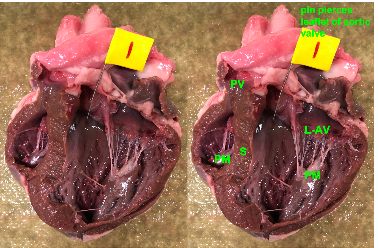
The image below shows a different heart that is dissected to show
the aortic valve. An unlabeled photo on the left allows you
to fully see the structures. PV: pulmonary
valve. PM: papillary muscle. L-AV: left AV
valve (mitral valve). S: interventricular
septum. Although not labeled, on these figures you should
also be able to identify the chordae tendineae and the left
atrium.
Video 5.1.6 ("Ventricles: outflow pathways") is
particularly useful for showing the structure and action of the
valves. The action of the valves is shown by pumping water through
the heart. When they are open, the valve cusps fall back against
the tissue walls. When the valves close, the edges of the cusps
come together. When viewed from the top, the three cusps of a
closed semilunar valve look like the Mercedes Benz logo.
What to identify in video 5.1.6 (opens in a new
tab):
ascending aorta
aortic valve
pulmonary trunk
pulmonary valve
pulmonary artery
AV valve
chordae tendineae
papillary muscle