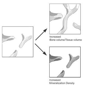

Bone mineral density increases when more mineral is packed within the bone, even if there has been no new bone formation (in fact, this may occur because there is so little bone formation). The mineralization density can be measured on bone samples from which marrow has been removed. The volume of bone is measured by fluid displacement and the mineral content determined by weighing the ashed specimen. Microradiograph and backscattered electron imaging are newer methods to measure mineralization density. This should not be confused with BMD or "bone mineral density" as measured by DEXA; the radiographic measurements cannot differentiate between increased bone volume and increased mineralization density, as shown in this figure:
Increasing mineralization density changes biomechanical properties of bone, making it stiffer but more brittle. Click to review basic .
The optimum mineralization density in human bone is unknown, but patients with osteoporosis have lower mineralization density than patients with normal bone, and treatment with bisphosphonates increases the mineralization density. This increase in mineralization density accounts for most of the increase in DEXA measurements in people treated with bisphosphonates. Click to see an animation of bone treated with bisphosphonates, which increase mineralization density.
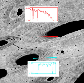
| Electron backscattered image of iliac cortical bone showing two osteons with plot profiles taken along the thick lines. The red profile shows that density is low in the newly formed osteoid (dark gray) and gradually increases. The blue profile shows a more abrupt change from the mineralized bone to the Haversian canal space; this is an area of bone resorption. The downward spikes in the plot profiles represent the osteocyte lacunae. |