SKELETAL MUSCLE PHOTOS - PART 2 MAMMALIA (EUTHERIA) - Cat |
|
Check out the variation between the different cats dissections. Click on each image to see the larger version.
Lateral Views showing gluteals & biceps femoris; anterior is to the right. Lateral Views - fascia & muscle of tensor fascia latae has been removed to expose the vastus lateralis.
Medial Views of superficial muscles, sartorius & gracilis Medial View
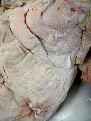
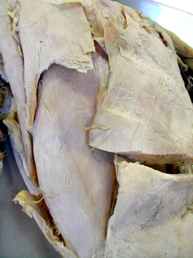

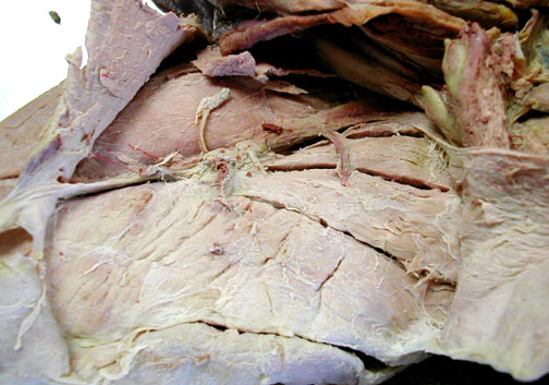
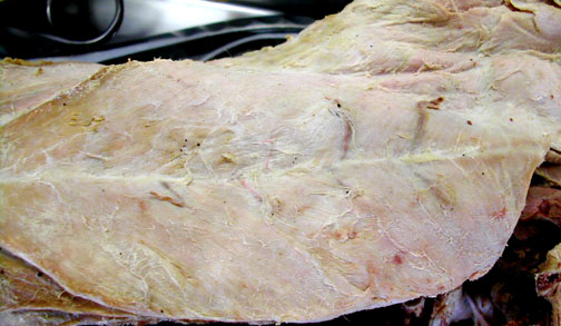
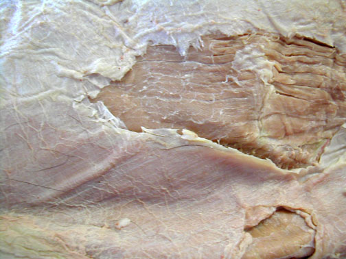
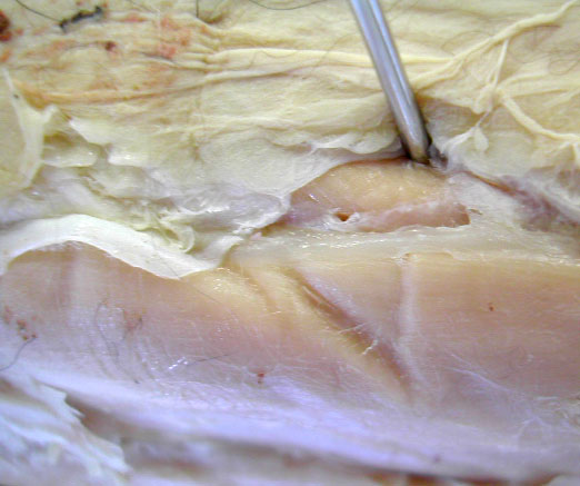
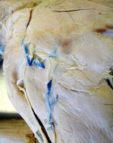
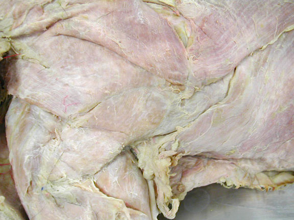
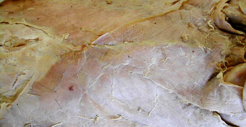
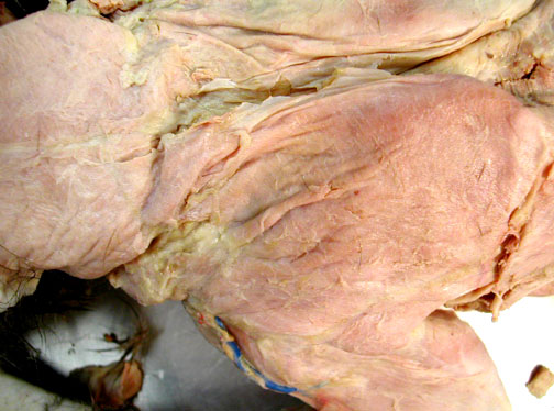
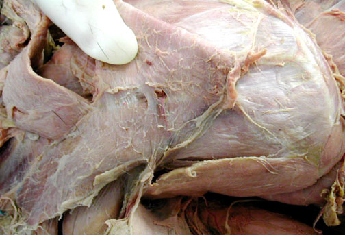
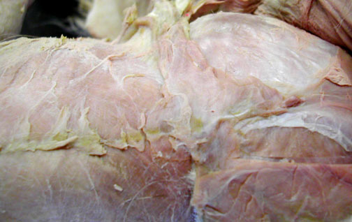
Pectoralis Muscles on ventral side of chest |
|
Head to the right
|
Head to the left
|
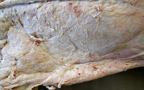
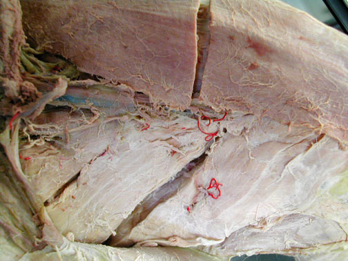
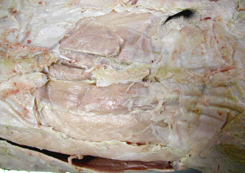
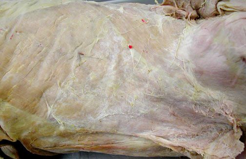
External oblique & linea alba
|
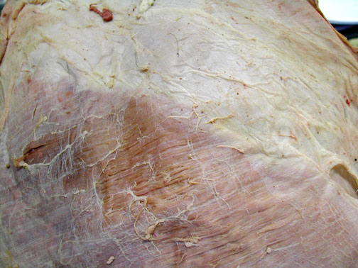
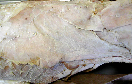
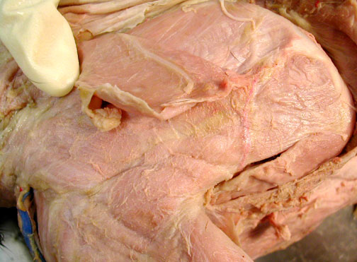
Head is to the left, with exposed rectus abdominus muscle fibers by removing part of the linea alba
|
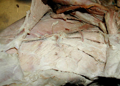
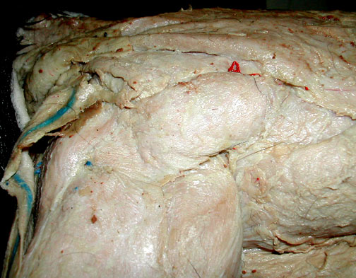
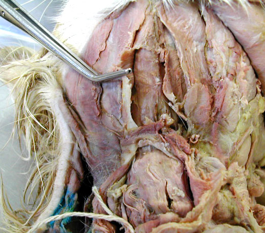
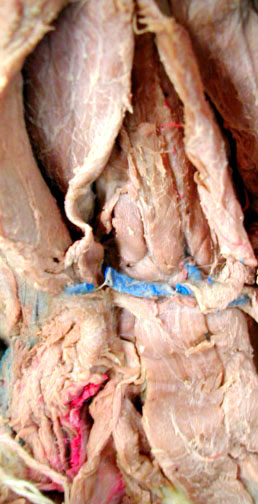
Dorsal - Lumbar Region |
|
Head is to the left; the lumbodorsal fascia has been removed, in part to expose the large bundles of the longissimus dorsi & the more medial multifidis spinae |
|
|
|
Shoulder |
|
|

|
 |
 |
Lateral or Dorsal Views - superficial; head to the right; latissimus dorsi & spinotrapezius visible |
|
 |
 |
Head is to the left in this specimen
|
Dorsal view
|
 |
Dorsal View - superficial head towards the top; clavotrapezius & acromiotrapezius visible.
|
Shoulder Dorsal Views; Deep - the acromiotrapezius has been peeled back to expose rhomboideus & serratus ventralis |
|
|
|
Serratus Ventralis - from a lateral view, very deep; latissimus dorsi has been removed, shoulder pulled out to expose posterior part of serratus ventralis. Head towards the left.
|
|
Lateral Views - deep; head to the left; acromiotrapezius partially removed to expose supraspinatus, infraspinatus & teres major |
|
 |
 |

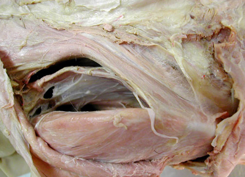

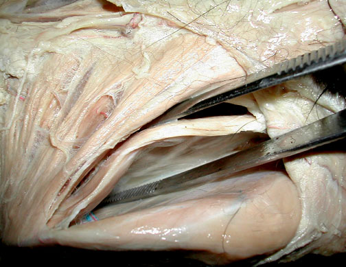
Throat & neck - anterior is towards the top of the screen |
|||
Ventral view; mylohyoid uncut, & digastric, sternohyoid visible
|
Ventral views; mylohyoid opened to expose the small geniohyoid, digastric & sternohyoid visible
|
||

Lateral view - far lower right shows the masster; a few lymph nodes & salivary glands are also visible
|
Dorsal view of the head, (facing to the right). Cutaneous muscles were cut to expose the temporalis muscle on the skull.
|

| top of page |