MRI
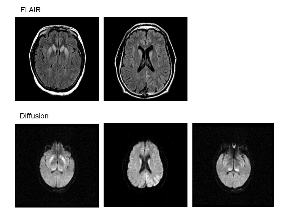
Findings: Axial flair and diffusion weighted (DWI) images showing hyperintensity in two areas. The left parietal cortex (not involving the subcortical white matter). Deep grey nuclei including the caudate, globus pallidus and thalamus. Gadolinium scans do not show enhancement (not shown). The apparant diffusion coefficient (ADC) is negative (not shown), which means that there is no ischemia.