 |
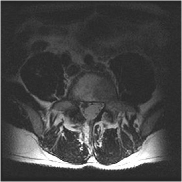 |
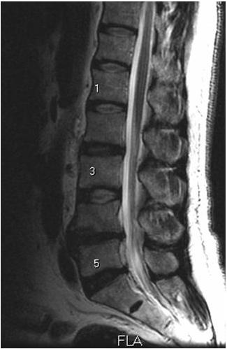
Midline sagittal, and L4-5 axial T2 images.
1. Degenerative changes involving osteophyte and disc bulging at L4-5 and
L5-S1.
2. No central spinal canal stenosis.
3. Right lateral disc herniation (arrow) displacing nerve roots posteriorly.