
Vertebral fractures
Other fractures
Changes in body shape
|
Hip Fractures Vertebral fractures Other fractures Changes in body shape |
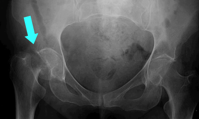
The arrow on this X-ray points
to a femoral neck fracture.
Hip fractures are a major cause of loss of independence in older women and men. Many are discharged into nursing homes instead of back to their previous living situation. The one-year mortality following a hip fracture is 12 to 24%. However, many of the patients who break their hip were frail and would be expected to have a high mortality rate anyway. It is estimated that 14% of deaths following a hip or pelvic fracture in previously ambulatory women were caused or hastened by the fracture.
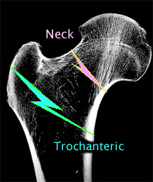 Hip fractures do not involve the hip joint itself, but are fractures of the proximal femur. About half of hip fractures are trochanteric and the others are femoral neck fractures. In older women the proportion of trochanteric fractures increases. Once age is taken into account, trochanteric fractures are more related to bone density, while femoral neck fractures are more related to mechanical factors. Here's an outside link to images of trochanteric fractures.
The vast majority of hip fractures occur after a fall. About 5% appear to be "spontaneous" fractures, in which the patient feels a fracture and then falls. Falls to the side are more likely to result in a hip fracture than forward falls.
Today most hip fractures are treated surgically, with pins, screws, or a hip replacement.
 Click on this cross-section of 3 lumbar vertebra to see how a compression fracture of the spine develops.
Notice that the spinal cord (yellow) is not compressed, and it is very rare for the "posterior elements" to fracture due to osteoporosis. With vertebroplasty, in which liquid plastic is injected into the compressed vertebral body to restore height, there is about a 5% incidence of serious leakage of the plastic into the spinal cord which can cause spinal cord damage.
About 60% of women with compression fractures |
Vertebral compression fractures vary in degree from mild wedges to complete compression. The symptoms also vary, but the degree of compression is not necessarily related to the amount of pain. It is possible that some of the fractures occurred gradually and therefore did not cause acute pain. Nevitt took spine xrays in 7223 older women and repeated the xrays 3.7 years later. During that time 371 women had a new vertebral fracture. Women were asked carefully about pain and disability symptoms. Increased back pain was experienced in 22% of women who did not have a new fracture and 38% of women who did have a new fracture.
These findings are similar to those in clinical trials, in which new vertebral fractures are found by xrays but only a minority of patients were aware of the occurance of the fracture. I think this has several implications for the management of osteoporosis. Since vertebral fractures strongly predict future fractures, and since many of them are "silent", it makes sense to recommend spine xrays as part of the evaluation of patients at risk for osteoporosis.
When women and men do suffer painful compression fractures, the pain usually lasts from one to two months, is localized to the back with accompanying muscle spasms, then gradually subsides. Patients with continuing severe pain should be evaluated for other pathologic etiologies of the fracture, especially malignancy or myeloma. Persistent pain can also be caused by continuing fracture, muscle spasms, spinal stenosis, or degenerative joint disease.

To correctly interpret a spine xray, it is important to know the definition of a vertebral fracture, which is not quite as straightforward as it first appears, especially for research. For practical clinical purposes, a vertebra can be considered fractured if the anterior height is 80% or less of the posterior height. A new fracture requires loss of at least 20% of anterior or posterior height.
This illustration is from Genant J Bone Mineral Res 1996;11:986 |
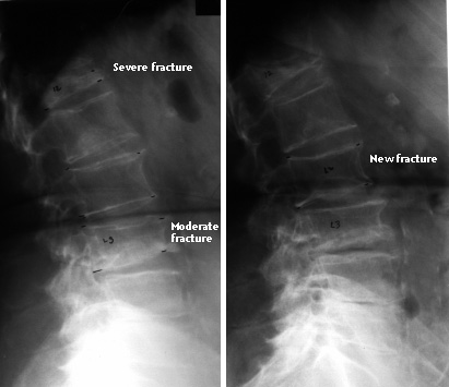 Here is an xray from a patient who had a severe fracture at T12 and a moderate fracture at L3. Three years later a second xray revealed a new fracture at L2. |
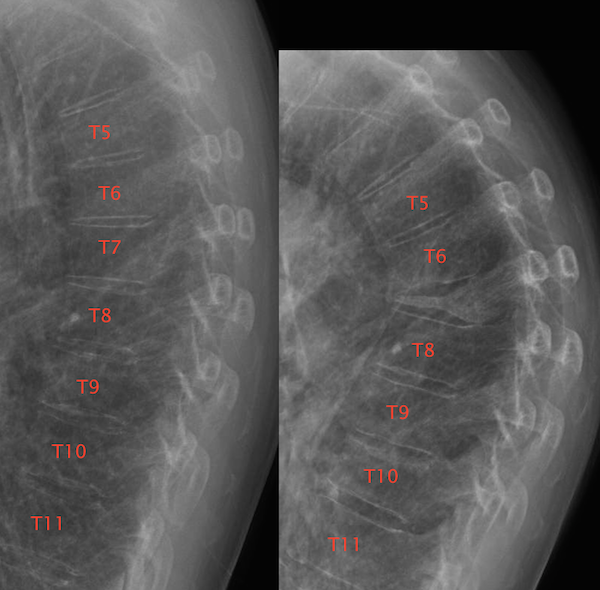
This is another before and after image of a severe fracture at T7 and moderate fracture at T10.
Wrist fractures (Colles fractures) are more common in women who are 50 to 60 years old. These are caused by falls or other trauma. Osteoporosis does not appear to impair the healing of the wrist fractures, and they cause only short-term disability.
Although spine, hip, and wrist fractures are considered classical osteoporotic fractures, many others are related to bone density and thus are also osteoporotic. These include rib, pelvic and shoulder fractures, but not finger, facial bone, skull, elbow, or ankle fractures. More details about which fractures are osteoporotic are on the page about fracture risk factors.
Click on the drawing to see how vertebral compression fractures cause changes in posture and body shape. |
 |

Click on picture above to see a Quicktime movie (900Kb) This is a clip from a 20/20 show broadcast 20 years ago and just as true today! KYPHOSIS The "Dowager's hump" caused by vertebral compression fractures is disfiguring. This is the feature of osteoporosis that is identified by most patients. The hump causes difficulty in finding clothes that will fit, let alone look attractive. In severe cases, the ribs contact the iliac crest and movement causes pain. HEIGHT LOSS The irreversible height loss associated with osteoporosis is one of the aspects of the disease that is most distressing to many women. They have trouble reaching high shelves, driving the car, and are at greater risk for airbag injuries. Height loss can also occur with scoliosis, which often gets worse after menopause. Also, degenerative disk disease can cause height loss of 2 inches. Some reversible height loss is due to poor posture. |
PROTRUDING ABDOMEN The protruding abdomen which is a result of the kyphosis is an unrecognized aspect of osteoporosis. Women do not realize that the curvature of the spine decreases the abdominal space, and thus the intestines have nowhere to go except forwards. Many women think that they are getting fat, and they go on a diet trying to regain their youthful waistline. If they do successfully lose weight, it will only increase their risk for more osteoporotic fractures. It is important for the physician to explain to the patients that the protruding abdomen is a consequence of kyphosis, and that other women with osteoporosis also really dislike this feature of the disease. Also, some fashions can minimize the appearance of the kyphosis and the protruding abdomen.
DECREASED PULMONARY CAPACITY Patients with kyphosis have decreased lung volumes. In severe cases this leads to shortness of breath and pulmonary symptoms of restrictive lung disease.
REFLUX ESOPHAGITIS Patients with kyphosis may develop reflux esophagitis due to the changes in abdominal space. Wearing tight clothing can exacerbate the problem.
Updated 3/14/2015, reviewed 2/10/16