
Pulmonary Function Testing
Case 4 Answer
A 25 year-old man presents to his physician with complaints of dyspnea and wheezing. He is a non-smoker. Two years ago, he was in a major motor vehicle accident and was hospitalized for 3 months. He had a tracheostomy placed because he remained on the ventilator for a total of 7 weeks. His tracheostomy was removed 2 months after his discharge from the hospital.
His pulmonary tests are as follows:
| Pre-Bronchodilator (BD) | |||
|---|---|---|---|
| Test | Actual | Predicted | % Predicted |
| FVC (L) | 4.73 | 4.35 | 109 |
| FEV1 (L) | 2.56 | 3.69 | 69 |
| FEV1/FVC (%) | 54 | 85 | |
| Check abbreviations>> | |||
His flow volume loop is as follows:
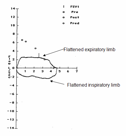
This patient has evidence of airflow obstruction on spirometry as he has a low FEV1 and a reduced FEV1/FVC ratio of 0.54. Given that the FEV1 is 69% of predicted this patient would be labeled as having “mild airflow obstruction.
In order to make a correct diagnosis in this patient, however, you cannot look simply at the numbers from his spirometry testing but must also look at the flow volume loops. A noteworthy feature of his flow volume loop is that there is flattening of both the inspiratory and expiratory limbs. This pattern is seen in patients who have a fixed upper airway obstruction. In a patient with a prior history of tracheostomy, you would be very suspicious that this patient has developed tracheal stenosis, a known long-term complication of tracheostomy tubes.
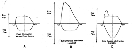
Other forms of airway obstruction will also demonstrate characteristic patterns on the flow-volume loops. Patients with a variable intrathoracic obstruction (eg. a carcinoid tumor in a mainstem bronchus) have flattening of the expiratory limb of the flow-volume loop while patients with variable extrathoracic obstruction (eg. a thyroid tumor) have flattening of the inspiratory limb of the flow-volume loop.
All three of these patterns are demonstrated in the figure below.
UW School of Medicine : School of Medicine Mission
Copyright and Disclaimer : Credits and Acknowledgements