
Pulmonary Function Testing
Case 9 Answer
A 35 year-old previously healthy man presents with dyspnea, fevers, chills and night sweats for the past 2 months. He is a non-smoker with no concerning habits or occupational exposures. His pulmonary function tests are as follows:
Her pulmonary function testing is as follows:
| Pre-Bronchodilator (BD) | |||
|---|---|---|---|
| Test | Actual | Predicted | % Predicted |
| FVC (L) | 1.66 | 4.48 | 37 |
| FEV1 (L) | 0.94 | 3.67 | 26 |
| FEV1/FVC (%) | 57 | 82 | |
| RV (L) | 1.39 | 1.66 | 84 |
| TLC (L) | 3.06 | 5.96 | 51 |
| RV/TLC (%) | 45 | 29 | |
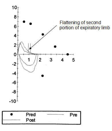
His flow volume loops is as follows:
Case 9 Interpretation
This patient has reduced FEV1 and FVC with a low FEV1/FVC ratio, consistent with an obstructive process. He also has a low TLC indicating he has a restrictive process as well. He would, therefore, be labeled as having a combined obstructive-restrictive defect. His obstructive defect would be classified as “very severe” based on his FEV1 of only 26% predicted while his restrictive process would be labeled as moderate given his TLC of 51% predicted.
There is an interesting finding in his flow-volume loop as well. The expiratory limb appears to have two components. There is a steep component and then a second, flatter component over the latter half of exhalation. This pattern suggests that one lung may be emptying faster than the other and, therefore, that the slowly emptying lung might have an obstructing airway lesion.
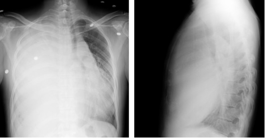
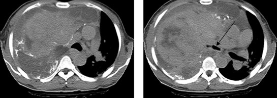
In fact, when the patient underwent chest x-ray imaging, he was found to have a dense opacity in the right chest. CT scanning revealed the presence of a large mass. This mass was so large it not only collapsed the right lung but also compressed the left lung causing lung restriction. It also compresses the airways (arrow in CT scan) on the right side leading to the obstruction to air-flow out of the viable right lung. These images are shown below.
Chest X-ray
Chest CT
UW School of Medicine : School of Medicine Mission
Copyright and Disclaimer : Credits and Acknowledgements