
A Primer on Reading Pulmonary Function Tests
by Joshua Benditt, MD
Print primer [220k PDF*]
- What are PFTs used for?
- What types of measurements can be made in PFT?
- What determines the major lung volumes?
- What determines airflow through the bronchial system?
- How to interpret PFTs
- An algorithm for interpreting PFTs
- How to report PFTs
What Are Pulmonary Function Tests Used For?
Pulmonary function testing provides a method for objectively assessing the function of the respiratory system. The tests do not always diagnose specific conditions but should be used to gain a greater understanding of a patients' clinical problem. Questions which may be answered with pulmonary function tests include:
- Identification of certain primary diseases of the respiratory system.
- Following the course of a specific disease over time.
- Quantitation of the severity of disease.
- Assessment of a response of a disease process to treatment.
- Exclusion of certain disease processes from diagnostic consideration (e.g. upper airway obstruction).
Pulmonary function tests must always be analyzed within the context of the patient being tested. Age, height, weight, race, and sex directly affect the results which one would predict for a given individual. Diseases which the patient may have or drugs which they are taking may be important in the interpretation of the patient's test. For instance, in a patient taking gold shots for rheumatoid arthritis, the finding of a restrictive PFTs, particularly if they are new, is very significant. Thus, the clinical context is extremely important in both understanding and interpreting PFTs.
What Types of Measurements Can Be Made In Pulmonary Function Testing
There are essentially four categories of information which can be obtained with routine pulmonary function testing:
- Lung volumes which can allow us to measure the maximum volume of the lungs as well as sub-compartments thereof.
- Flow rates which measure the maximal flow of gas out of (and sometimes into) the lung.
- Diffusing capacity which measures the transfer of gas from the alveolar space into the capillary blood stream.
- Maximal inspiratory and expiratory pressures which measure the applied strength of the respiratory muscles.
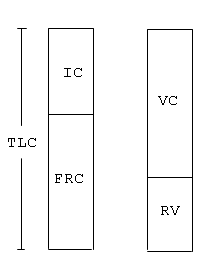
Prior to examining how each of the measurements are made, let us examine some of the volumes and flow rates which we will be using in our evaluation of PFTs. Although the lung volumes can be divided into a large number of compartments including volumes and capacities (which are the combination of two or more volumes), there are four important volumes which should be remembered:
- total lung capacity (TLC) or the total volume of gas contained in the lungs;
- functional residual capacity (FRC) or the volume of gas left in the lungs with the individual relaxed at the end of expiration;
- residual volume (RV) the volume of gas left in the lungs at the end of forced expiration; and
- vital capacity (VC) the difference between the largest (TLC) and the smallest (RV) lung volumes which can be obtained. These volumes are shown in Figure 1.
Fig 1
Measurements of Lung Volumes
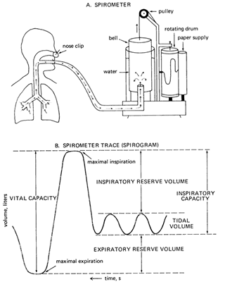
Measurement of some of the volumes such as vital capacity is easy and can be performed with the simple spirogram. By having the patient breath to their maximal capacity (TLC) lung capacity and blow out as far as possible (RV), the vital capacity can be recorded (see Figure 2 below).
Fig 2
Other volumes such as residual volume (RV) and total lung capacity (TLC) cannot be measured with the spirometer but require an additional measurement technique, either the body plethysmograph or helium dilution in order to be determined. This is because the amount of gas left in the thorax at maximal expiration (RV) cannot be measured by the spirometer. By using one of the other techniques, we can determine this volume and subsequently all other volumes and capacities including TLC. (The body plethysmograph and helium dilution techniques are shown in Fig 3a below).
Fig 3a
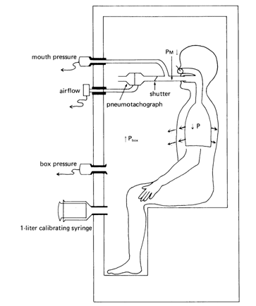
Pbox x Vbox = Pbox f x (Vbox i -∆V)
Once ![]() V has been solved for we can then go on to solve for the thoracic gas volume in the following equation:
V has been solved for we can then go on to solve for the thoracic gas volume in the following equation:
PMi x VLi = PMf x (VLi + ∆V)
This equation follows from the Boyle's Law and tells us that the initial pressure measured at the mouth (PMi) times the lung volume at which that pressure is measured (VLi) will be equal to the new mouth pressure (PMf) x the new lung volume (VLi + ∆V) while the patient is making small changes in their lung volume by panting against the closed shutter.
Fig 3b
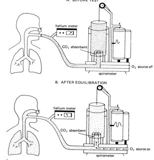
The helium-dilution technique makes use of the following relationship: If the total amount of substance dissolved in a volume is known and its concentration can be measured, the volume in which it is dissolved can be determined.
Amount of solute = concentration of solute x volume of solvent
In the helium-dilution technique, helium is inspired and dissolved in the gas in the lungs. The concentration of helium is determined with a helium meter. allowing calculation of the patient lung volume. Helium is used for this test because it is not taken up by the pulmonary capillary blood. The total amount of helium does not change during the test. The helium concentration is monitored continuously with a helium meter until its concentration in the inspired air equals its concentration in the subject's expired air. At that point the concentration of helium is uniform in the spirometer and the patient's lung.
The test is stopped at the end of a normal tidal volume, FRC and the volume of FRC is calculated:
Initial Concentration of helium x Initial Spirometer Volume =
Final Concentration of Helium x (Final Spirometer Volume + FRC)
Fhei x Vsp = Fhef (Vspf + VLf)
What Determines the Major Lung Volumes?
Total lung capacity is determined by the ability of the inspiratory pump (brain, nerves, muscle) to expand the chest wall and lungs which have a strong tendency to recoil inwards at high lung volumes. Any breakdown in the ability of pump to function will result in a smaller total lung capacity (restrictive lung disease). Neuromuscular disease is an example of this. Diseases which increase inward recoil of the lung (pulmonary fibrosis) will lead to a smaller TLC. Diseases which lead to a reduction in inward recoil of the lung (emphysema) result in an increase in TLC known as hyperinflation.
Residual volume (RV) is determined in healthy younger individuals by the competition between the strength of the expiratory muscles and compressibility of the chest wall. However, by the onset of middle age or in obstructive lung disease RV appears to be determined by a "flow limitation"; expiratory flow rates at low lung volumes are so low that expiration is prolonged and is not completed down to the original RV by the time the subject gives up the effort and takes another breath.
Vital capacity (VC) is determined by the difference between TLC and RV and changes with variations in RV or TLC. It is easily measured and reliable and can check the measured validity of a measured change in RV or TLC. FRC is the relaxation volume at the end of expiration. It is not a reliable measurement and requires excellent cooperation on the part of the subject. In patients with obstructive lung disease FRC may be elevated. This imposes a significant extra load on the inspiratory muscles which can results in muscle fatigue.
What Determines Airflow Through the Bronchial System?
Air flows through a tube if there is a pressure difference between the ends. In the respiratory system the pressure difference is between the alveolar pressure and the pressure at the airway opening or mouth. Flow may be laminar (smooth) or turbulent dependent on characteristics of the gas and the tube through which it is traveling. Most of the resistance to airflow occurs in the first few divisions of the airways. The more distal airway divisions, because of their large cross-sectional area, constitute a silent zone of airway resistance.
Resistance to flow is not constant at all lung volumes. As the lung expands, airways enlarge reducing the airways resistance at high lung volumes. Airways resistance increases at lower lung volumes. A plot of airways resistance vs. lung volume is shown in Fig 4.
Fig 4
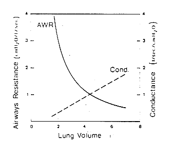
Other factors besides lung volume can affect airway resistance. Smooth muscle within the wall of the same bronchi can contract and increase airways resistance. Secretions in airways or edema in the airway wall can also increase airways resistance. In patients with emphysema, loss of tethering of small airways open during exhalation leads to collapse and an increase in resistance to airflow. All obstructive lung diseases are characterized by an increase in resistance to expiratory flow.
Measurement of Expiratory Flows
Measurement of expiratory flow is extremely useful to us particularly in identifying obstructive lung disease but in a number of other ways also. Expiratory flows are measured during the forced expiratory spirogram (Figure 2). Again, the patient breaths to TLC and forcefully exhales to residual volume generating the expiratory spirogram with volume plotted against time.
The spirogram can be broken up into subdivisions. The ones which we are most concerned about are
- the FVC which has been mentioned previously and represents the entire volume exhaled from the lungs in a forced breath,
- the FEV1 or the volume of gas exhaled in the first one second of exhalation,
- the FEF25-75 which is the flow of gas exhaled during the middle half of the vital capacity previously known as the maximal mid expiratory flow or (MMFR).
The forced expiratory maneuver has been called "an unnatural act" because it is rarely if ever performed during daily activities. Nevertheless, it probes a very important pathophysiologic limit. Beyond a modest expiratory effort, the limit to flow is effort-independent; pushing harder does absolutely no good. The limit, however, is markedly volume dependent ranging in healthy persons from 10 liters per second at high lung volumes to near zero flow at RV. The limit is lowered at all lung volumes by primary narrowing of airways or narrowing due to decrease in lung recoil (emphysema) and is responsible for the ventilatory impairment seen in these obstructive lung diseases.
Flow Volume Loop Analysis
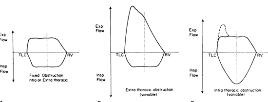
In addition to portraying the spirogram as volume plotted against time, it can also be plotted as flow against volume as shown below in figure 5. This can be particularly helpful in identifying obstruction lesions of the upper airway.
Fig 5
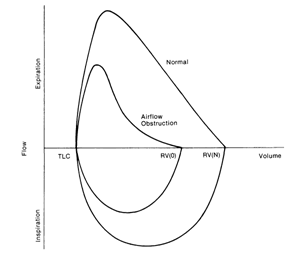
Fig 6: Intra and extrathoracic large airway obstructing lesions
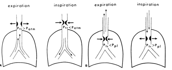
Fig 7: Flow-volume loops in intra and extrathoracic lesions
Diffusing Capacity
The diffusing capacity is a measure of the transport of gas across the alveolo-capillary membrane. The techniques of this measurement is discussed will be discussed with you. The diffusing capacity reflects the surface area of the alveolo-capillary membrane as well as its thickness and the driving pressure for gas across the membrane. It can be reduced in diseases such as emphysema, pulmonary fibrosis, or pulmonary vascular disease. It can also be reduced in patients with anemia.
How To Interpret Pulmonary Function Tests
One of the first questions in interpreting pulmonary function testing is the definition of what is "normal". A great deal of data has been amassed in an attempt to determine what is normal for an individual of a given height, race, sex, and age. Despite the large amount of data gathered, many questions and interpretation problems still exist. However, we must do the best job with the data we have available. Currently, the most commonly used method of deciding whether a measured value falls outside of the normal range is to take the measured value for that individual and compare it with a mean value measured for a group of similar individuals. If the individual's value falls outside of the predicted value by 20% or more, then it is said to be abnormal. For example, if an individual's TLC is predicted to be 8 liters (100%) and the measured value is 6 liters (75%), then this is an abnormally low value. Obviously values immediately around the "magic" 80% mark must be interpreted with caution and will need to be interpreted in the light of other measurements. Some authors use the concept of the 95% confidence interval for those values falling within the normal range.
The questions which we will be able to answer with a complete set of pulmonary function tests are:
- Is there obstruction to airflow present?
- If there is airway obstruction
- Is there a restrictive process present?
- Is it a parenchymal process?
- Is it an extraparenchymal process?
- Is the extraparenchymal process a neuromuscular problem?
- Is there a combined obstructive restrictive disorder present?
- Is there an isolated gas exchange abnormality?
- Is there upper airway obstruction present
- Is it variable or fixed and intra or extrathoracic?
1. Is Obstruction Present?
In all cases of obstruction there will be a reduction in expiratory flow as noted on the spirogram. The FEV1 will be reduced. However, this value might also be reduced in restrictive lung disease. How do we deal with this problem? Two strategies have been devised. It has been noted for some time that in obstructive lung disease, although all indices of flow decrease, the FEV1 tends to decrease more than the FVC. Thus in individuals with obstruction, the FEV1/FVC tends to be reduced to a value below that predicted for normal individuals. If one has only spirometric data available, the diagnosis of obstructive lung disease can be made by a finding of a reduction in the FEV1 and FEV1/FVC. The ATS has defined the lower limit of normal (LLN) for the FEV1/FVC as the predicted value for that individual – 9 for women and predicted value – 8 for men. Some athletes and older people will have an abnormally low FEV1/FVC ratio. This does not indicate an obstructive ventilatory defect. There is no reduction in FEV1.
If the full set of lung volumes has also been measured, then other clues to an obstructive process will be available. A very sensitive indicator of obstruction to airflow is an increase in the RV which has been referred to as airtrapping. With more severe obstruction to airflow, increases in FRC and TLC can also be seen. Thus the characteristic findings of an obstructive defect on pulmonary function testing include a reduction in FEV1, a reduction in the FEV1/FVC, and an increase in RV with either a normal or increased TLC. Occasionally, in mild obstructive lung disease, the only defect which may be seen is a reduction in FEF25-75.
2. How Severe is the Obstruction?
The severity of obstruction is graded on the basis of the reduction in FEV1 and has been determined by agreed on standards from the American Thoracic Society.
- FEV1 < 65-80 % mild obstruction
- FEV1 < 50-65% moderate obstruction
- FEV1 < 50% severe obstruction
3. Is the Obstruction Fixed or Reversible?
In some obstructive airways diseases, a part or all of the obstruction will be reversible with bronchodilators. Therefore in all cases where the technician notes obstruction, two inhalations of a bronchodilator will be given to the subject. An improvement of 12% in the FEV1 or FVC is considered a significant response with an increase of at least 200ml. Asthma is considered the prototypical disease reactive to bronchodilators. However, more "fixed" types of obstruction such as emphysema and chronic bronchitis may also show reversibility. In addition, because asthma is a variable disease, at times pulmonary function tests may appear entirely normal. One will therefore make the diagnosis by clinical history or attempt to provoke obstruction using a "bronchoprovocational" agent such as methacholine or cold air which can illicit bronchoconstriction which might not otherwise be seen.
4. Is the Disease Consistent with Emphysema?
Emphysema is a diagnosis made by the pathologist examining lung tissue and now more recently with a typical pattern on thoracic CT scan. However, there are certain findings on pulmonary function testing which can point towards a diagnosis of emphysema. A reduction in FEV1, FEV1/FVC as well as an increase in RV are seen. The TLC is elevated consistent with a reduction in inward elastic recoil of the lung because of destruction of elastic tissue. Frequently, a reduction in DLCO reflecting destruction of the alveolo-capillary bed is also seen. The flow-volume loop may also show findings of dynamic airway collapse.
5. Is Restrictive Lung Disease Present?
The defining factor for restrictive lung disease is the reduction in the TLC. TLC, RV, VC, and FRC all tend to be reduced, though not in all cases. Measurements of expiratory flow tend to be preserved including the FEV1/FVC and FEF25-75.
6. What Type of Restrictive Pattern is Present?
Parenchymal processes result in a restrictive pattern by reducing the compliance or "stretchability" of the lung. Frequently in these processes there is a destruction of the alveolo-capillary bed which is seen as a reduction in the DLCO. A reduction in the TLC coupled with a reduction the DLCO points to a parenchymal cause of restrictive disease.
Diseases outside of the lung which prevent maximal expansion of the respiratory system including neuromuscular, skeletal, and even extrathoracic processes such as ascites or pleural effusion can lead to restrictive ventilatory defects. A neuromuscular disease such as Duchenne's muscular dystrophy affects the muscles of expanding the chest wall. All lung volumes will be reduced in a nearly proportionate way. The DLCO will usually be normal because there is no intrinsic problem with the lungs. One lung volume, expiratory reserve volume (ERV) may actually be greater than predicted because of weak expiratory muscles. The finding of a reduction in maximal inspiratory and expiratory pressures confirms the cause of restrictive defect.
Abnormalities in the skeletal system or chest wall itself can result in a restrictive ventilatory defect. The kyphoscoliosis can result in reductions in TLC with a preserved DLCO as can such unusual entities such as fibrothorax, massive ascites, or obesity. In these cases muscle strength and DLCO may appear normal.
How Severe is the Restrictive Defect?
Based on American Thoracic Society criteria, restrictive lung disease is based on the criteria of TLC.
| 65-80% | mild restriction |
| 50-65% | moderate restriction |
| < 50% | severe |
7. Is there a combined disorder (obstructive and restrictive) present?
On occasion there can be a combination of obstruction and restrictive processes occurring simultaneously. For instance, a patient who smokes and has developed emphysema and later presents with a neuromuscular cause of restrictive lung disease. Some diseases can intrinsically have both a restrictive and an obstructive component such as sarcoidoisis in which there may be an endobronchial component as well as an interstitial component causing restrictive lung disease. In these cases, the finding will be a combination of a reduction of TLC associated with reduction in flow, namely a decrease in FEV1 and FEV1/FVC ratio.
8. Is there an isolated gas exchange abnormality present?
Sometimes the only abnormality noted on pulmonary function testing is a reduction in DLCO. This test is quite variable and difficult to perform so that in general concern is not raised until the DLCO is approximately 60% or less than that of predicted. Isolated reductions in DLCO may be an early sign of interstitial lung disease, a vasculitis, pulmonary emboli, or anemia. The DLCO can be corrected for anemia to rule out the latter.
9. Is Upper Airway Obstruction Present?
Upper airway obstruction may be suggested by the clinical findings of stridor on physical examination. Reductions in flow are usually seen on the forced expiratory maneuver. However, when flow is plotted against volume evidence of upper airway obstruction can be readily appreciated. Abnormalities in the flow volume cure are immediately appreciated. Intra and extrathoracic variable and fixed lesions can be lesions can be identified, ranging from mediastinal tumor to an enlarged thyroid. (See figure 5 below Q: is this fig 5 above or another fig?)
An Algorithm for Interpreting PFTs
Question 1: Obstruction
Is there obstruction to airflow present?
| Men | Lower limit of FEV1/FVC | < Predicted- 8% |
| Women | Lower limit of FEV1/FVC | < Predicted- 9% |
How severe is it?
| Mild Obstruction | FEV1 65 - 80% predicted |
| Moderate Obstruction | FEV1 50 -65% predicted |
| Severe Obstruction | FEV1 < 50% predicted |
Is it reversible or fixed?
- Does the FEV1 or FVC increase by at least 15% after inhalation of a bronchodilator?
Is it possibly consistent with emphysema?
- Are lung volumes increased consistent with air-trapping
- Is the DLCO reduced consistent with loss of alveolocapillary membrane
Question 2: Restriction
Is there a restrictive process present?
- TLC less than 80% predicted
How severe is it?
| Mild | TLC or FVC | 65 - 80% predicted |
| Moderate | TLC or FVC | 50 -65% predicted |
| Severe | TLC or FVC | < 50% predicted |
Is it a parenchymal process?
- Reduction in DLCO is consistent with parenchymal destruction
Is it an extra-parenchymal process (kyphosis, muscle weakness)?
- DLCO will usually be normal.
- Maximal Inspiratory and expiratory pressures reduced
Is there a combined obstructive restrictive disorder present ?
- TLC is low and FEV1/FVC ratio is low
Question 3: Combined Obstructive and Restrictive Disease
Concomitant reduction in TLC and FEV1/FVC
Quantitation of severity
- Obstruction:Use FEV1
- Restriction: Use TLC or VC
Examples
- Sarcoidosisis, CF, obliterative bronchiolitis
- COPD + muscle weakness
Question 4: Isolated Pulmonary Vascular Disease
Is there an isolated gas exchange abnormality?
- Normal PFT’s other than reduction in DLCO
- Think of:
- Pulmonary embolism
- Pulmonary vascular disease – (e.g.,, pulmonary artery hypertension)
- Early interstitial lung disease
How To Report PFTs
The report includes
- the tabulation of results of the tests performed, juxtaposed with the predicted values for the subject, generated by the technician and
- an analysis and
- summary generated by me.
I attempt to keep the report short. The longer, the less likely to be read. In the analysis, I do not repeat the findings except as significant positives or negatives and I always state them in the context of the analysis. For example, "The increase in the RV and the decrease in the indices of forced expiratory flow and the specific airways conductance indicate obstructive airways disease."
I attempt to make the logic explicit. For example, "The decrease in TLC indicates restriction. The markedly diminished MIP suggests that this is due to chest wall disease while the normal diffusing capacity suggests that it is not due to a parenchymal process, such as interstitial fibrosis". This keeps me intellectually honest, and communicates more meaningfully.
Prior tests can be very valuable because comparison with self is inherently more sensitive than comparison with population norms and may give essential information about the progress of the disease or the positive or negative response to treatment. I always look at all the previous results. I often select out specific items for tabulation (my secretaries are very good at pulling out the numbers in the finished report if I simply say "please make a table showing the TLCs, the VCs, and the DLCOs for all of those tests") when progression is worth reviewing. I do, however, analyze the findings in the current test on its own merits before turning to comparison with previous tests, which, I suspect, has on occasion kept me from propagating a prejudice.
The Summary gives the major conclusions including qualifications, important outstanding questions, and suggestions for how one might proceed. It is brief (shorter than the analysis) and does not repeat the findings or the logic. It is intended to tell the referring physician what I think is going on and to help him or her to decide what to do.
First, I decide what my bottom line is going to be and how to qualify it. For example, "Moderate restrictive process probably due to a parenchymal disease, with an independent obstructive component."
Second, I try to envision what this report will do for the referring physician. The physician may have posed a particular question such as "Preop for bronchogenic carcinoma" which warrants a specific comment. If the referring physician has questioned asthma and is not in a subspecialty that handles asthma often, I may say "These findings do not rule out the clinical diagnosis of asthma". In an extremely obese patient who has perfectly normal pulmonary function tests, obstructive sleep apnea and obesity hypoventilation spring to mind and should be mentioned. If pulmonary fibrosis is suspected, I may suggest that "if clinically indicated, we could probe the possibility of gas exchange abnormality more finely with oximetry, arterial blood gases, and steady state diffusing capacity during rest and exercise". If a test result is very surprising or potentially urgent (a preoperative patient, or a PaO2 of 43), I contact the physician directly by phone!
UW School of Medicine : School of Medicine Mission
Copyright and Disclaimer : Credits and Acknowledgements