Pleural Effusions
Case 10 Answers
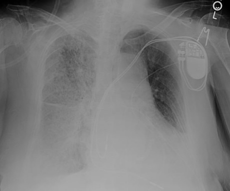
A 55 year-old man presents with increasing dyspnea and is found to have a very large right pleural effusion occupying half of his chest cavity on chest x-ray. A thoracentesis is performed and 2.5 liters of serosanguinous fluid is removed. He initially feels like his breathing is markedly improved and the x-ray shows that you drained out most of the fluid in the pleural space. While you are awaiting the results of the pleural fluid studies, however, the patient develops increasing dyspnea at rest and his oxygen saturation falls to 85% on room air. You obtain a chest x-ray, which shows the following:

How would you interpret the chest x-ray on this patient?
The patient has diffuse right lung opacities and a small residual right pleural effusion
What is the etiology of this increasing dyspnea and hypoxemia?
If too much pleural fluid is drained from the pleural space in one thoracentesis, patients are at risk for developing a non-cardiogenic form of pulmonary edema known as re-expansion pulmonary edema. This phenomenon can also be seen after overly rapid reexpansion of a collapsed lung in pneumothorax. Patients develop hypoxemia and dyspnea. Because this is due to a capillary leak phenomenon, patients may also demonstrate hemodynamic instability. Edema typically develops on the side of the drainage procedure but can occur bilaterally. It typically comes on within the first several hours of draining the fluid but can occur up to 24 hours later. The risk of reexpansion pulmonary edema is greatest when there has been overly rapid expansion of a chronically collapsed lung (more than several days).
How could this have been prevented?
Because the risk of this phenomenon is highest with overly rapid expansion of a collapsed lung, this complication could have been prevented by limiting the volume of removed fluid. Only 1 to 1.5L of fluid should have been removed during the procedure.
What is the appropriate management at this time?
Treatment of reexpansion pulmonary edema is largely supportive. There is no role for diuretic therapy as the patients are often intravascularly volume depleted since this is a capillary leak phenomenon. Fluids and pressors may be necessary to support patients with low blood pressure. Patients with severe hypoxemia may require CPAP therapy while in other situations mechanical ventilation is necessary. Recovery usually occurs spontaneously within 1-2 days of the event.
UW School of Medicine : School of Medicine Mission
Copyright and Disclaimer : Credits and Acknowledgements