
Pleural Effusions
Case 6
(This case is courtesy of Leo Bennett, MD, Madigan Army Medical Center.)
A 37 year-old woman presents with 10-days of progressive exertional dyspnea. Her review of systems was pretty unremarkable. She denied any fevers, chills, cough, sputum production or weight loss. The only recent event that she can recall was that she had an upper respiratory tract infection 3 months ago at the same time that multiple family members were sick. Her symptoms resolved without antibiotic therapy. Her only past medical history is allergic rhinitis. She is a life-long non-smoker and had a recent negative mammogram. A chest x-ray is performed and is shown below:
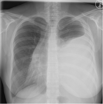
How would you interpret her chest x-ray?
Thoracentesis is performed revealing straw-colored fluid with 2875 RBC, 448 WBC (differential 18% polymorphonuclear cells, 37% lymphocytes, 35% macrophages and 10% mesothelial cells). The gram’s stain is negative. LDH 406 (serum value 180), total protein 4.7 (serum value 7.0). Cytology is performed and no malignant cells are seen.
How would you interpret the results of these pleural fluid studies?
A CT scan of the chest is performed. Two representative images are shown below.
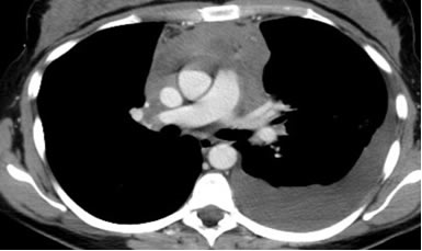
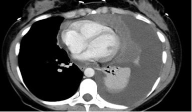
What are the noteworthy findings on the CT scan? What is the differential diagnosis for these findings?
What further diagnostic steps are warranted?
UW School of Medicine : School of Medicine Mission
Copyright and Disclaimer : Credits and Acknowledgements