
Pleural Effusions
Case 4
A 34 year-old woman with a history of heavy alcohol use presents to the emergency room complaining of increasing shortness of breath and right-sided chest pain. Her past medical history is remarkable for several prior episodes of pancreatitis, likely secondary to her chronic alcohol use. A chest x-ray is performed and is shown below:
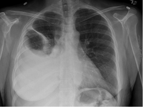
She initially undergoes thoracentesis for symptom relief but studies are not performed on the fluid. A repeat thoracentesis is then performed and 2 liters of serosanguinous fluid are removed. The pleural fluid studies reveal LDH 894 (serum value 175), total protein 3.6 (serum value 5.3), WBC 4000 with 90% eosinophils. The gram’s stain is negative.
How would you interpret the results of the pleural fluid analysis?
What do you make of the high percentage of eosinophils in the pleural fluid?
What additional tests should you order on the pleural fluid?
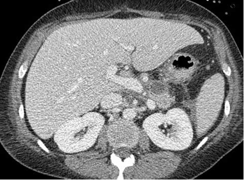
A CT scan of her chest and abdomen is performed. The scan reveals the presence of a possible pancreatic cyst, shown below. This prompts the physicians to order a pleural fluid amylase, which comes back at 4855 (normal < 20). How does this change your differential diagnosis?
How can you distinguish between pancreatic and other causes of amylase-rich pleural effusions?
What can you do to manage this patient’s pleural effusion?
UW School of Medicine : School of Medicine Mission
Copyright and Disclaimer : Credits and Acknowledgements